Skin surface electrodes

a technology of skin surface electrodes and electrodes, which is applied in the field of measuring or detecting electrical signals, can solve the problems of high cancer mortality rate, difficult to achieve the effect of reducing air pressure and reducing air pressur

Inactive Publication Date: 2012-10-25
EPI SCI LLC
View PDF9 Cites 36 Cited by
  • Summary
  • Abstract
  • Description
  • Claims
  • Application Information

AI Technical Summary

Benefits of technology

[0008]In another embodiment, an apparatus for determining electrophysiological characteristics of subepithelial tissue, the apparatus comprising a cup adapted to contact epithelial tissue comprising a surface having a concave shape, having a volume enclosed by the cup and the epithelial tissue, the cup capable of maintaining a reduced air pressure and holding a volume of flowable material; and an electrical support structure comprising a connection between an instrument for measuring or recording electrical signals and a plurality o

Problems solved by technology

Difficulty in detecting abnormal pre-cancerous or cancerous tissue before treatment options become non-viable is one of the reasons for the high mortality rate from cancer.
Detecting the presence of abnormal or cancerous tissues is difficult, particularly where affected tissues are located beneath the skin surface, for example, deep within the body, thus requiring expensive, complex, invasive, and/or uncomfortable procedures.
Thus, the use of detection procedures is often restricted or delayed until a patient experiences symptoms related to abnormal tissue.
Many forms of cancers or tumors, however, require extended periods of time to attain a detectable size and thus to produce significant symptoms or indicate their presence in the patient.
It is often too late for effective treatment by the time detection is performed with currently available diag

Method used


Image

  • Skin surface electrodes
  • Skin surface electrodes
  • Skin surface electrodes

Examples


first embodiment

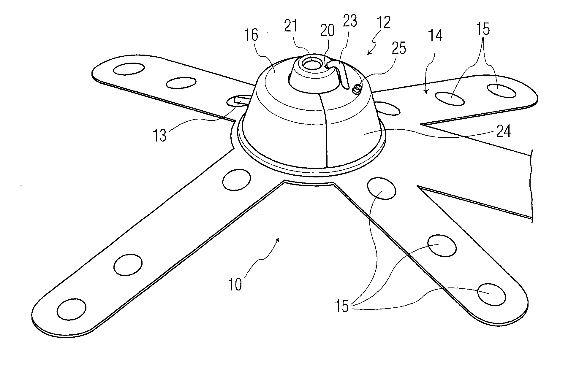
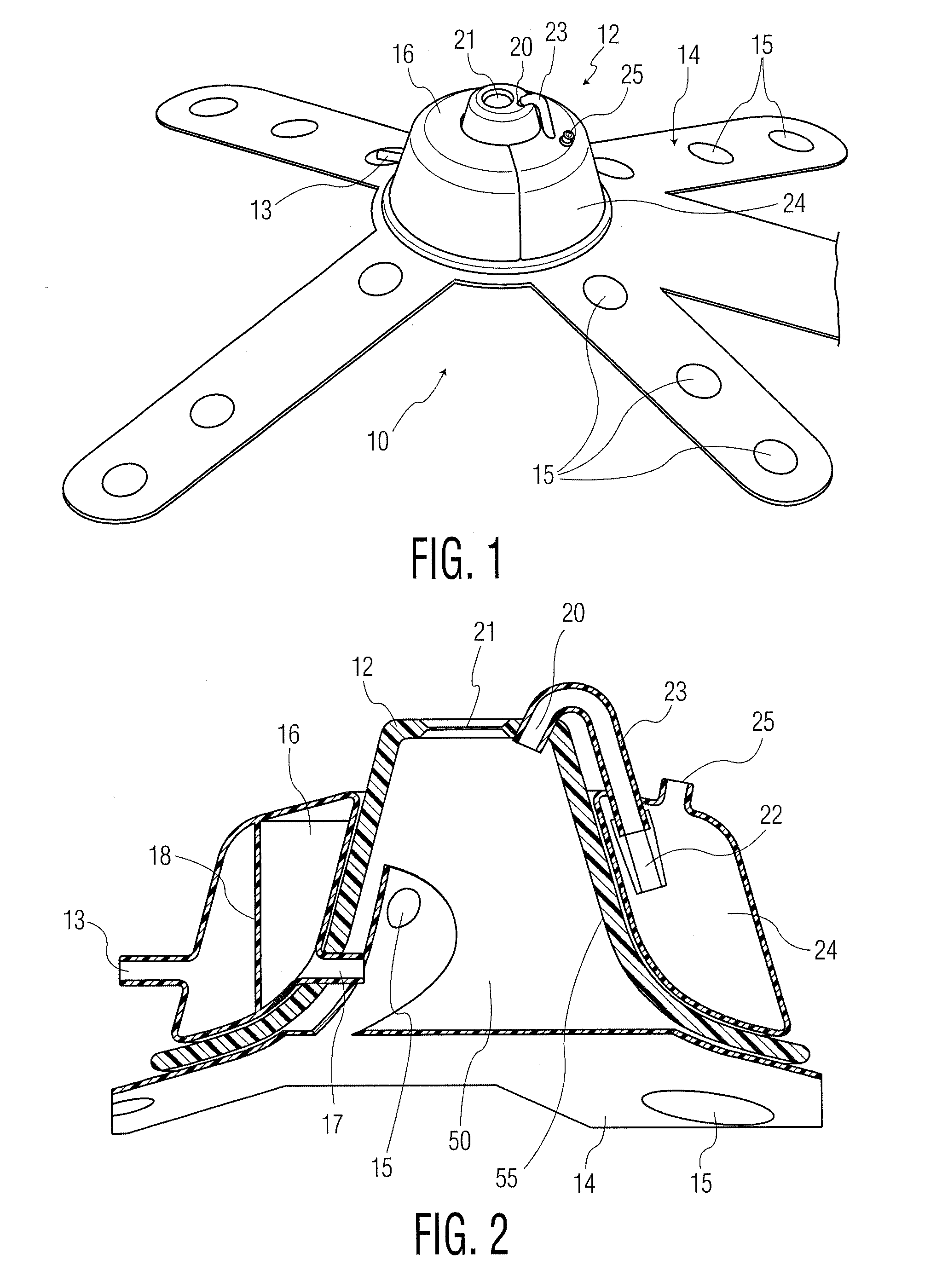
[0022]The present invention overcomes deficiencies associated with prior devices. In a first embodiment, as exemplified in FIG. 1, the apparatus 10 of the present invention includes generally a cup 12 and an electrical support structure 14. Apparatus 10 may be positioned on an epithelial tissue, such as the skin and / or nipple of a breast, and may be used to send and / or receive electrical signals through the epithelial tissue to measure, for example, density, of subepithelial tissue which may be tissue under the surface tissue (e.g., skin, nipple surface, etc.) and may include breast tissue, epithelial ductal tissue, deep tissue, and the like.

[0023]As illustrated in FIG. 2, the cup 12 may have an open volume 50 which is bounded by the inner surface 55 of cup 12. The cup 12 may have any required shape, such as conical, half spherical, or the like, and may further have a domed tip (which may assist in collecting any remaining air within volume 50 during the process of reducing air pres...

second embodiment

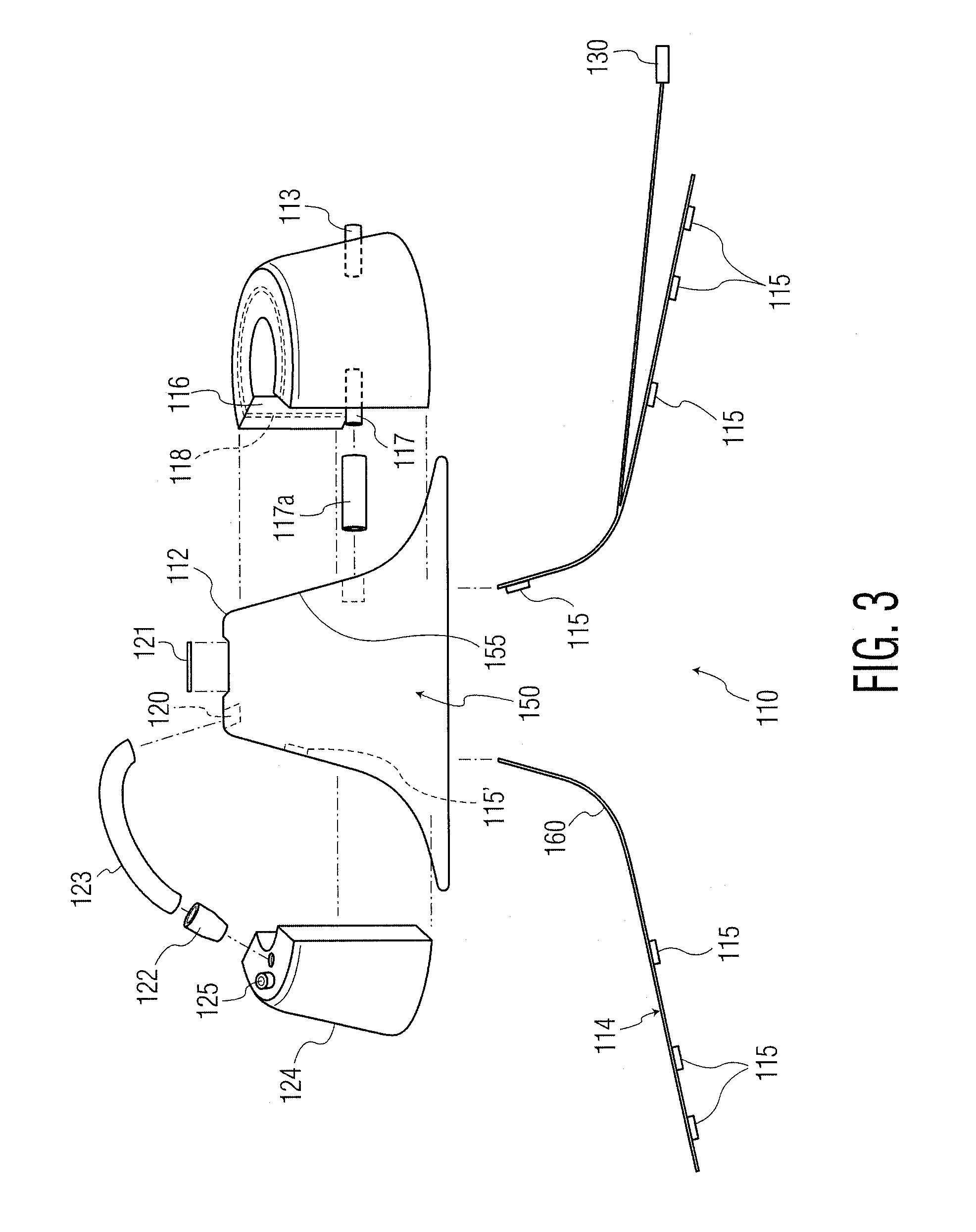
[0030]FIG. 3 illustrates an exploded view of apparatus 110 which may include cup 112, reservoir 116, a plurality of sensors 115 (as part of electrical support structure 114 or off the support structure 114, as with sensor 115′), exhaust / overflow reservoir 124 and at least two ports—fill port 117 and exhaust port 120. In this embodiment, the reservoirs are illustrated to be separate structures from the cup 112, and as such the reservoirs may be positioned anywhere relative to the surface 55 of cup 12. In FIG. 3, the reservoirs 116 and 124 may be connected by passageways 117a and 123, respectively. The exemplary embodiment of FIG. 3 may also include such elements as puncture seal 121, one-way valve 122, air exit port 125, reservoir liner diaphragm 118, pump port 113 (and pump, not shown), and the like. The surface 155 of cup 112, bounding volume 150, may further include a covering of at least a portion of surface 155 of adhesive foam 160. Adhesive foam 160 may be any material which pr...


PUM


Abstract

An apparatus (10) adapted to contact epithelial tissue, the apparatus comprising a cup (12) having a concave shape and adapted to be positioned on epithelial tissue, the cup capable of maintaining a reduced air pressure and holding a volume of flowable material; and an electrical support structure (14) comprising a support structure and a plurality of sensors, electrodes, or both (15) configured to interact with epithelial and subepithelial tissue.

Description

CROSS-REFERENCE TO RELATED APPLICATIONS[0001]The present application claims the benefit of Application Ser. No. 61 / 279,471, filed Oct. 21, 2009, entitled SKIN SURFACE ELECTORDES, the disclosure of which is hereby incorporated herein by reference.BACKGROUND OF THE INVENTION[0002]The present invention relates generally to the measurement or detection of electrical signals such as in abnormal or cancerous tissue, and more particularly, to the detection of changes in the electrophysiological characteristics of abnormal or cancerous tissue and to changes in those electrophysiological characteristics related to the functional, structural and topographic (the interaction of shape, position and function) relationships of the tissue during the development of malignancy. Measurements can be made in the absence or presence of pharmacological, hormonal or other chemical agents to reveal and accentuate the electrophysiological characteristics of abnormal or cancerous tissue.[0003]Difficulty in d...

Claims


Application Information

Patent Timeline
IPC IPC(8): A61B5/053
CPCA61B5/053A61B5/4312A61B2562/043A61B2562/0209A61B5/6834A61B2562/0217A61B2562/0215
Inventor DAVIES, RICHARD J.DAVIES, MARY K.
Owner EPI SCI LLC
Why Eureka
Try Eureka