Method and apparatus for processing ultrasound image

Active Publication Date: 2013-01-17
SAMSUNG ELECTRONICS CO LTD
View PDF1 Cites 17 Cited by
  • Summary
  • Abstract
  • Description
  • Claims
  • Application Information

AI Technical Summary

Benefits of technology

[0014]The wavelet transformation may divide the reference ultrasound image into a plurality of band images in different frequency bands, and may be repeatedly performed with respect to a band image in a low frequency band to divide the r

Problems solved by technology

However, the diagnostic ultrasound imaging equipment provides a lower image quality than other medica

Method used


Image

  • Method and apparatus for processing ultrasound image
  • Method and apparatus for processing ultrasound image
  • Method and apparatus for processing ultrasound image

Examples


Embodiment Construction

[0045]The following detailed description is provided to assist the reader in gaining a comprehensive understanding of the methods, apparatuses, and / or systems described herein. However, various changes, modifications, and equivalents of the systems, apparatuses and / or methods described herein will be apparent to one of ordinary skill in the art. Also, descriptions of functions and constructions that are well known to one of ordinary skill in the art may be omitted for increased clarity and conciseness.

[0046]Throughout the drawings and the detailed description, the same reference numerals refer to the same elements. The drawings may not be to scale, and the relative size, proportions, and depiction of elements in the drawings may be exaggerated for clarity, illustration, and convenience.

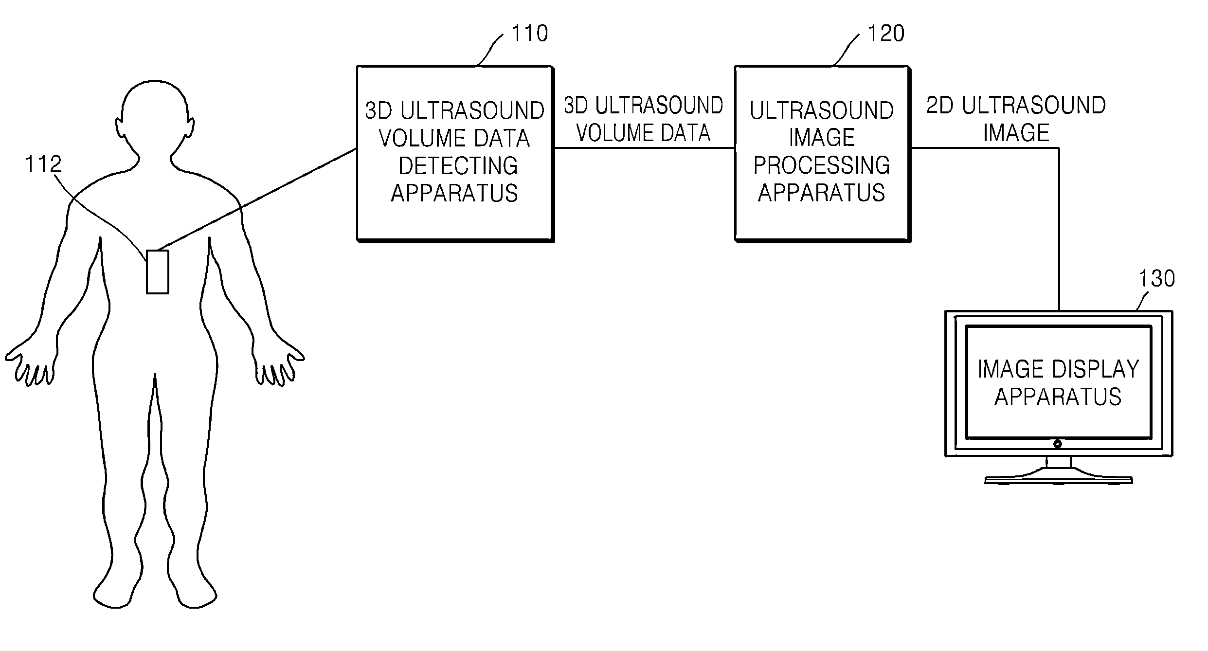
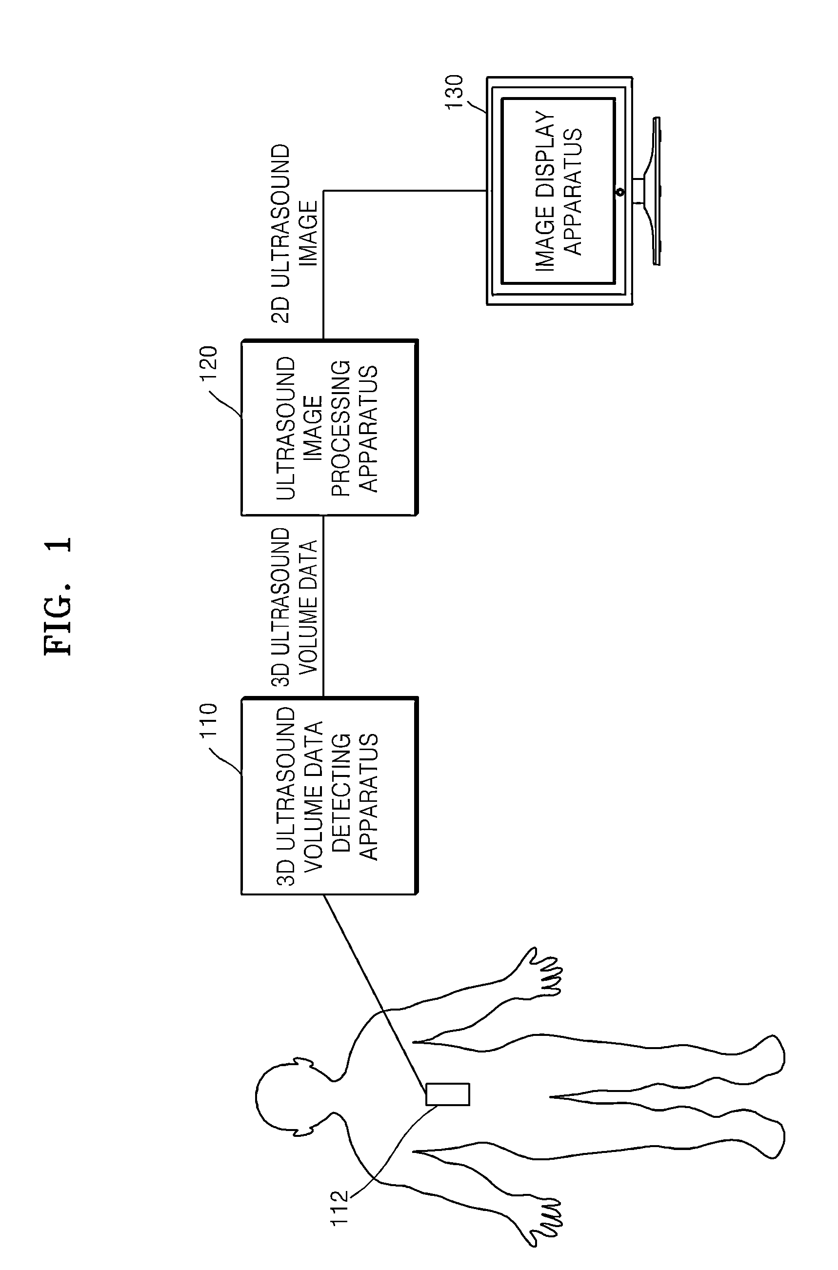
[0047]FIG. 1 is a schematic block diagram of an example of an ultrasound diagnosis system. Referring to FIG. 1, the ultrasound diagnosis system includes a three-dimensional (3D) ultrasound volume data...


PUM


Abstract

A method of processing an ultrasound image includes generating a plurality of two-dimensional (2D) ultrasound images from three-dimensional (3D) ultrasound volume data of an object to be diagnosed, generating a plurality of tissue edge images of an edge of at least one tissue component in the object to be diagnosed based on values of a plurality of pixels forming each of the 2D ultrasound images generated from the 3D ultrasound volume data, and generating a 2D ultrasound image from which a noise component has been removed by discriminating the edge of the at least one tissue component from a position of the noise component based on a difference between a similarity of the edge of the at least one tissue component in the tissue edge images and a similarity of the noise component in the tissue edge images.

Description

CROSS-REFERENCE TO RELATED APPLICATIONS[0001]This application claims the benefit of Korean Patent Application No. 10-2011-0068554 filed on Jul. 11, 2011, in the Korean Intellectual Property Office, the disclosure of which is incorporated herein by reference in its entirety.BACKGROUND[0002]1. Field[0003]This disclosure relates to methods and apparatuses for processing an ultrasound image by acquiring a plurality of ultrasound images from three-dimensional ultrasound volume data and post-processing the acquired ultrasound images to clarify the ultrasound image.[0004]2. Description of Related Art[0005]Recently, diagnosis equipment using ultrasound images have been widely used. Diagnostic ultrasound imaging equipment forms and provides an image of an object to be diagnosed, e.g., an organ, by radiating ultrasonic waves onto the object to be diagnosed and detecting and processing ultrasonic waves reflected from the object to be diagnosed. The diagnostic ultrasound imaging equipment allow...

Claims


Application Information

Patent Timeline
IPC IPC(8): A61B8/13
CPCA61B8/145A61B8/483A61B8/5269G01S7/52077G06T5/002G06T2207/30004G06T2207/10136G06T2207/20021G06T2207/20064G06T2207/20192G06T5/50G06T17/10
Inventor KIM, YUN-TAEKIM, JUNG-HO
Owner SAMSUNG ELECTRONICS CO LTD
Why Eureka
Try Eureka