Quantitative Elastography with Tracked 2D Ultrasound Transducers

a 2d ultrasound and quantitative technology, applied in the field of medical imaging, can solve the problems of inability to achieve ideal measurement systems, inability to achieve mr imaging, and relatively slow imaging mode, and achieve the effect of improving the accuracy of mr elastography and improving the accuracy of mr imaging

Active Publication Date: 2017-01-26
THE UNIV OF BRITISH COLUMBIA
View PDF3 Cites 9 Cited by
  • Summary
  • Abstract
  • Description
  • Claims
  • Application Information

AI Technical Summary

Problems solved by technology

Traditional diagnostic methods have relied on the use of manual palpation to discriminate between healthy tissue and diseased regions because imaging methods were unavailable for detecting changes in mechanical properties.
However, such ideal measurements systems are currently infeasible, so most previous mentioned works exploit the steady state nature of the wave propagation to build up a representation through multiple measurements over several periods of the waves.
MR imaging is a relatively slow imaging modality, so MR elastography typically requires many minutes of acquisition time.
Ultrasound holds promise for faster acquisition yet it poses other challenges to overcome due to the pulse-echo nature of data acquisition and need for multiple pulses, which introduce time delays from both time of flight of the pulses and the delays between pulses.
More challenges arise from the desire to acquire data over a 3D volume of interest when using a conventional ultrasound transducer that acquires data from a single 2D cross-sectional plane for a given position of the transducer.
This makes it more challenging to use conventional inversion methods to calculate the mechanical properties of tissue from the acquired measurements.
As mentioned that method though does not provide the quantitative mechanical properties of tissue, but the relative strain.
The image quality provided with that method can also be compromised by the accumulation of de-correlation due to involuntary motion of the user in all five degrees of freedom that violate the assumptions of only axial compression of tissue.
It is also only capable of measuring the static mechanical properties, not the dynamic properties that require measurements over time.
It is often the case that the frequency of tissue motion exceeds the imaging speed of commercially available ultrasound machines.

Method used


Image

  • Quantitative Elastography with Tracked 2D Ultrasound Transducers
  • Quantitative Elastography with Tracked 2D Ultrasound Transducers
  • Quantitative Elastography with Tracked 2D Ultrasound Transducers

Examples


Embodiment Construction

[0050]Detailed descriptions of embodiment of the invention are provided herein. It is to be understood, however, that the present invention may be embodied in various forms. Therefore, the specific details disclosed herein are not to be interpreted as limiting, but rather as a representative basis for teaching one skilled in the art how to employ the present invention in virtually any detailed system, structure, or manner. The descriptions of the embodiment of the invention will be made in the publication by Caitlin Schneider, Ali Baghani, Robert Rohling, Septimiu E. Salcudean titled “Remote Ultrasound Palpation for Robotic Interventions using Absolute Elastography”, presented at the 15th International Conference on Medical Image Computing and Computer Assisted Intervention, Oct. 2, 2012, Springer LNCS 7510, pp. 42-49, the entirety of which is hereby incorporated by reference.

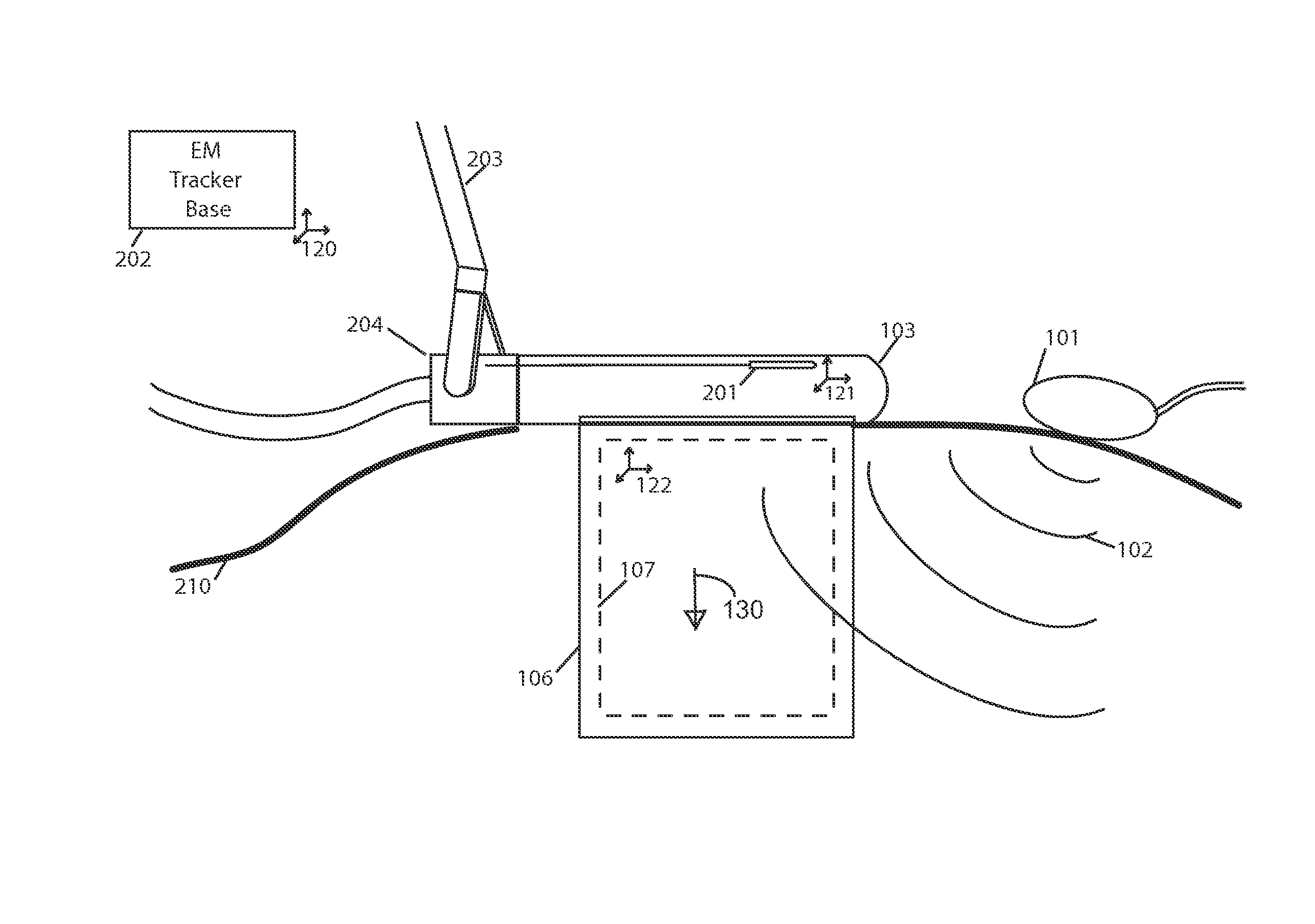
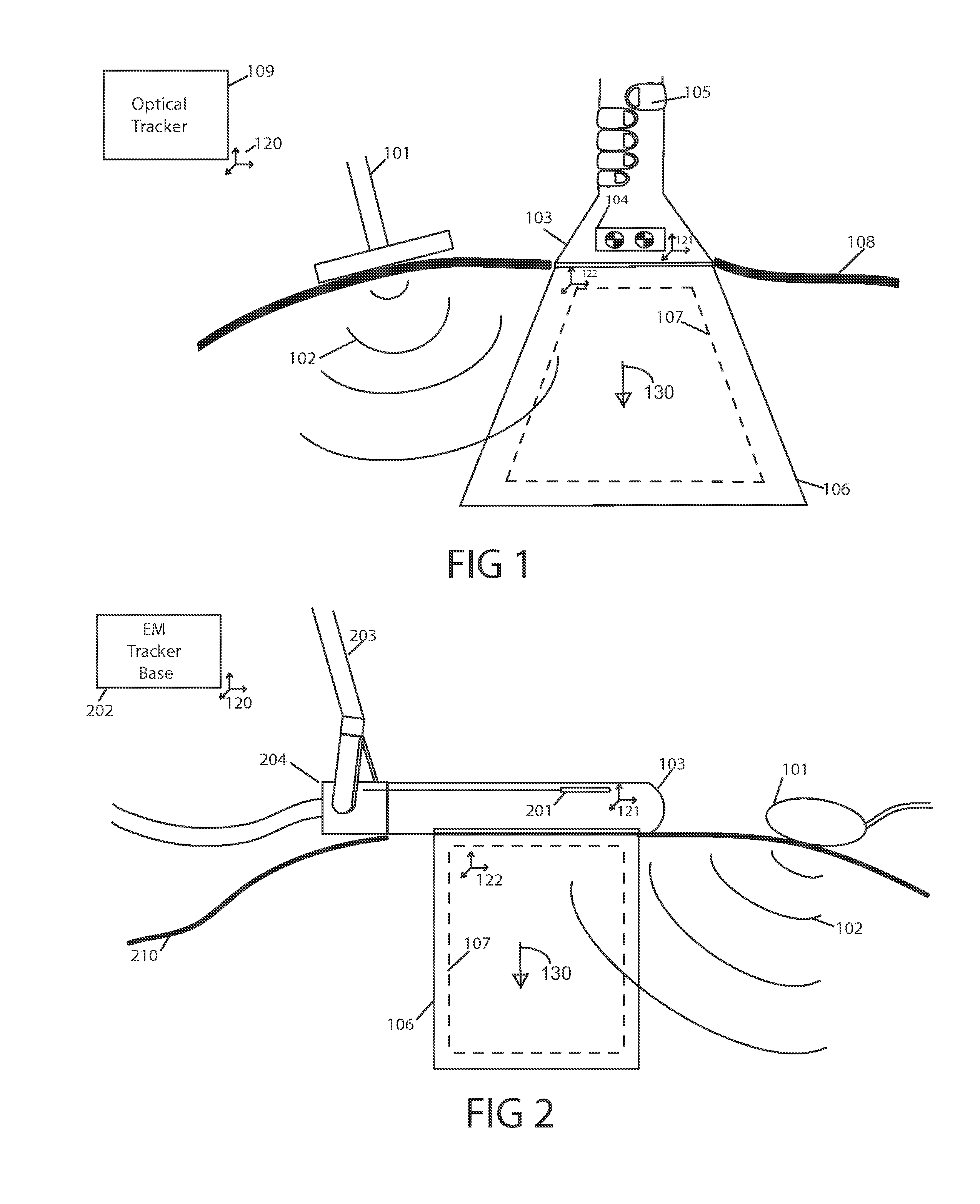
[0051]FIG. 1 shows one embodiment of the invention. In this embodiment, the ultrasound transducer 103 is bei...


PUM


Abstract

A method is described for acquiring 3D quantitative ultrasound elastography volumes. In one embodiment, the method comprises using a 2D ultrasound transducer to scan a volume of tissue through which shear waves are created using an external vibration source, the synchronized measurement of tissue motion within the plane of the ultrasound transducer with the measurement of the transducer location in space, the reconstruction of tissue displacements in time and space over a volume from this synchronized measurement, and the computation of one or several mechanical properties of tissue from this volumetric measurement of displacements. The tissue motion in the plane of the transducer may be measured at a high effective frame rate in the axial direction of the transducer, or in the axial and lateral directions of the transducer. The tissue displacements over the measured volume may be interpolated over a regular grid in order to make the computation of mechanical properties easier.

Description

[0001]This application claims priority under 35 U.S.C. §119(e) to a U.S. provisional application having Ser. No. 61 / 707,883, filed Sep. 28, 2012, the entirety of which is herein incorporated by reference.FIELD OF THE INVENTION[0002]This invention relates to medical imaging and, in particular, the measurement of mechanical properties of the tissues, also known as elastography.BACKGROUND OF THE INVENTION[0003]Medical imaging is used in many applications to determine the composition of tissue not visible to the naked eye. The images can be displayed to a user, where the intensity or color of the image is a function of some parameter of the tissue composition. For example, computed tomography (CT) displays to the user the absorption of X-rays in the body and ultrasound displays the echo pattern produced in response to a pulsed sound wave. Of particular interest are the mechanical properties of tissue which can also be depicted in images. Changes in the mechanical properties of certain t...

Claims


Application Information

Patent Timeline
Patent Type & Authority Applications(United States)
IPC IPC(8): A61B8/08A61B8/12A61B8/00
CPCA61B8/485A61B8/12A61B8/4263
Inventor SALCUDEAN, SEPTIMIU EDMUNDSCHNEIDER, CAITLIN MARIEROHLING, ROBERT N.BAGHANI, ALI
Owner THE UNIV OF BRITISH COLUMBIA
Why Eureka
Try Eureka