Ultrasound diagnostic and treatment device

a technology of ultrasound and diagnostic equipment, applied in the field of ultrasound equipment for diagnosis and treatment, can solve problems such as serious adverse effects, and achieve the effect of safe diagnosis and treatment of subjects

Inactive Publication Date: 2013-02-28
HITACHI LTD
View PDF19 Cites 25 Cited by
  • Summary
  • Abstract
  • Description
  • Claims
  • Application Information

AI Technical Summary

Benefits of technology

[0026]According to the present invention, a treatment using focused ultrasound can surely be performed in a short period, and from this effect, the present invention can provide a technique of safely diagnosing and treating a subject.

Problems solved by technology

In the HIFU therapy, spatial selectivity of therapy is achieved by the convergence of ultrasound alone, and therefore, the HIFU therapy has a possibility that, if the region is misaligned due to a body motion, high intensity ultrasound of 1 kW / cm2 or more is exposed to a region other than the region to be treated, which causes serious adverse effect.

Method used


Image

  • Ultrasound diagnostic and treatment device
  • Ultrasound diagnostic and treatment device
  • Ultrasound diagnostic and treatment device

Examples


first embodiment

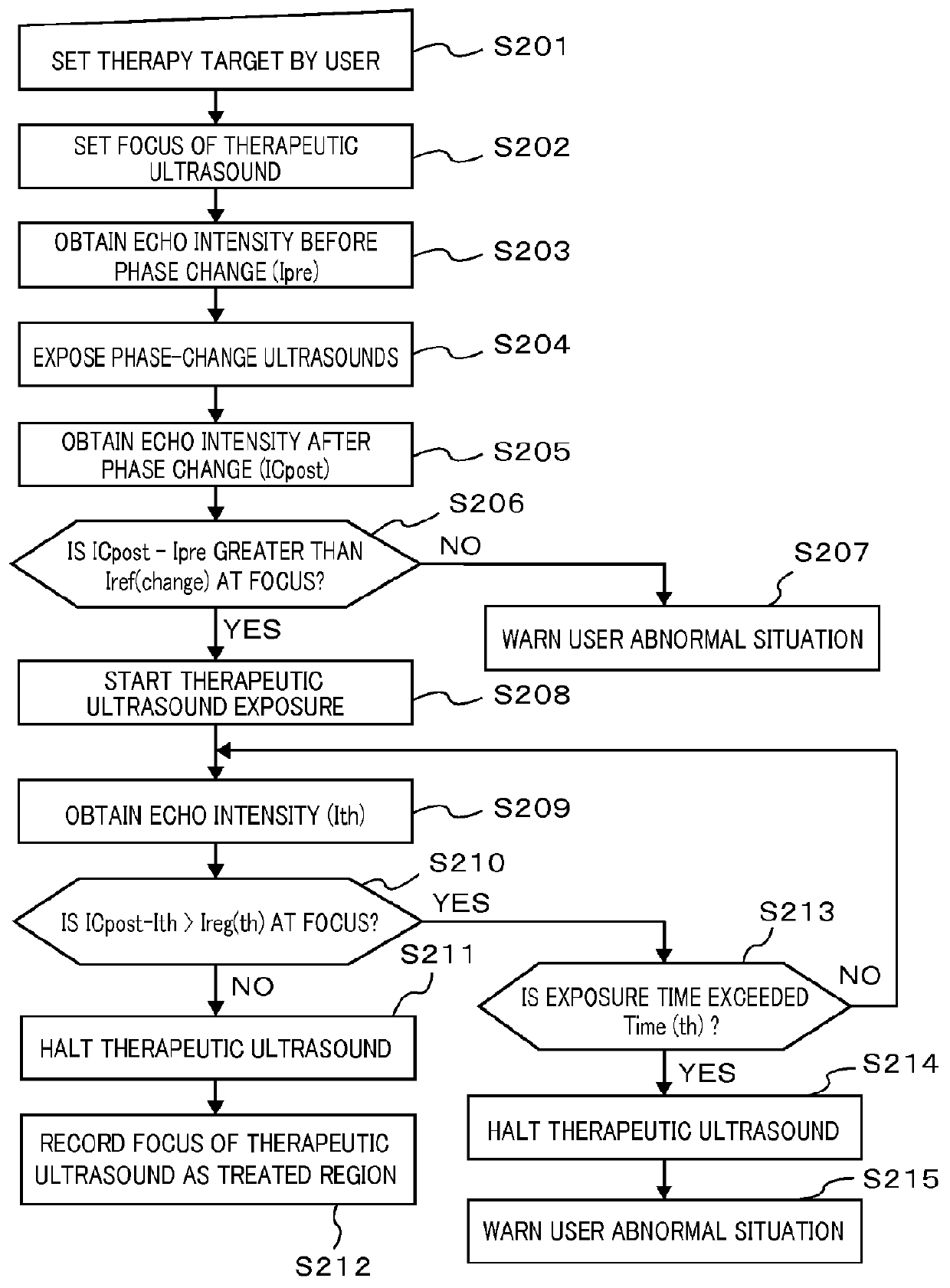
[0058]A configuration and an operation of a first ultrasound apparatus for diagnosis and therapy will be described based upon FIGS. 1 to 5, 12, and 13.

[0059]

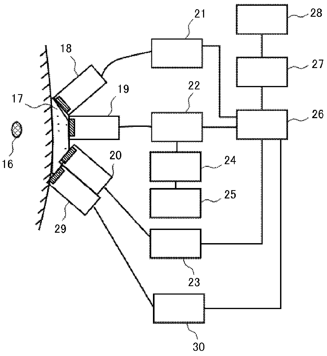
[0060]FIG. 1 is a view illustrating one example of a schematic configuration of an ultrasound apparatus for diagnosis and therapy according to the first embodiment. In this figure, the ultrasound apparatus for diagnosis and therapy includes a phase-change ultrasound transmit unit 18 that is arranged for a therapy target (region) 16 through an acoustic coupling media 17 for exposing phase-change ultrasound, an ultrasound transmit unit 29 for microbubble sustention, which generates ultrasound for sustaining the microbubbles generated by a phase change, an ultrasound receive section for phase change unit 19 that emits phase-change detection ultrasound to the therapy target 16, and receives the phase-change detection ultrasound reflected from the therapy target 16, and a therapeutic ultrasound transmit unit 20 for emitting therapeut...

second embodiment

[0095]

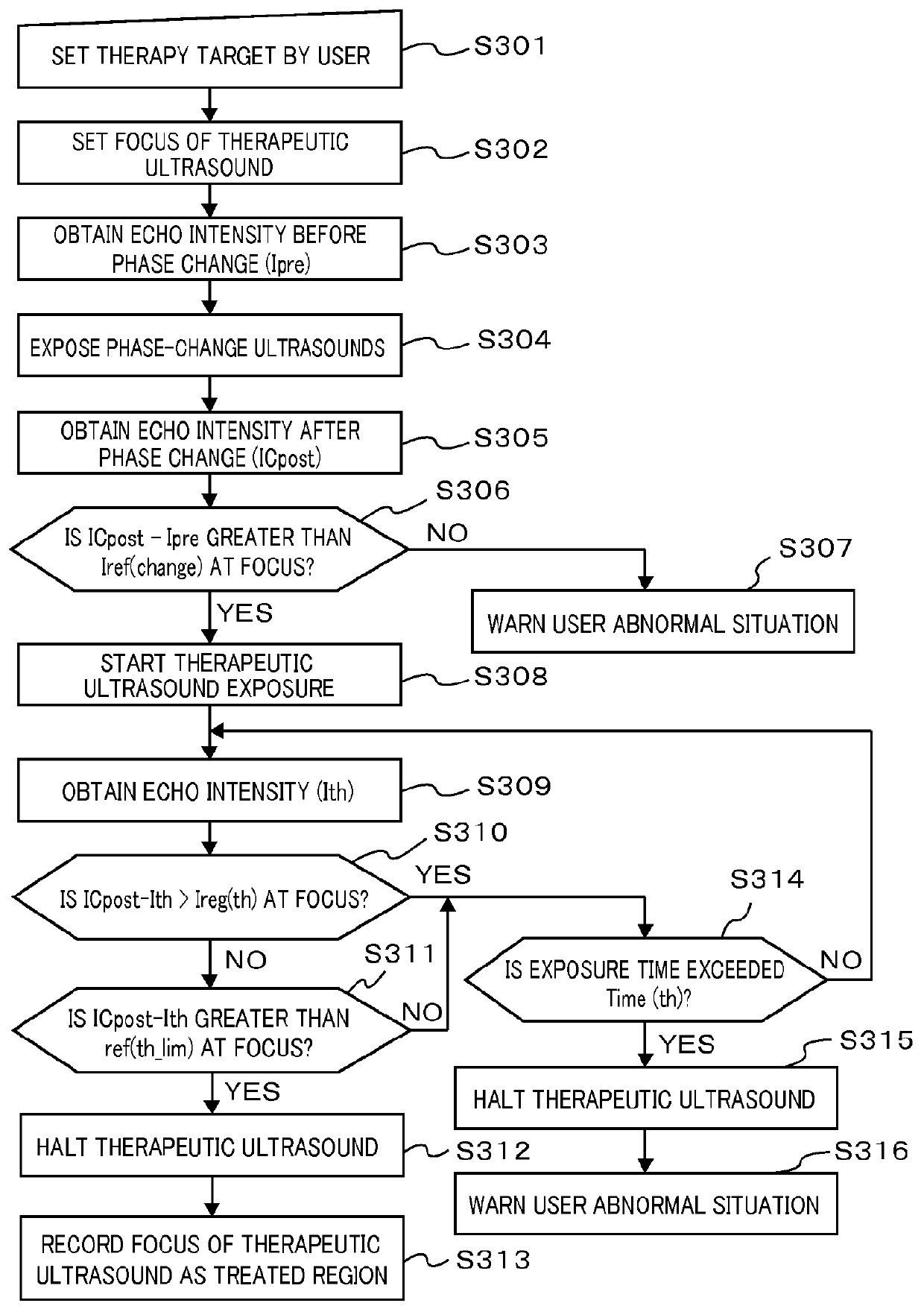
[0096]The ultrasound apparatus for therapy illustrated in FIG. 1 having the configuration described above transmits and receives four types of ultrasounds, which are the phase-change ultrasound, phase-change detection and confirmation ultrasound, microbubble sustention ultrasound, and therapeutic ultrasound. For the phase-change detection ultrasound, an ultrasound receive section having a function equivalent to the function of a probe for a normal ultrasound diagnostic device is exclusively used, but it is considered to be advantageous for simplifying the apparatus and securing safety by using the same ultrasound exposure apparatus for the other ultrasounds. A second embodiment in which a single ultrasound exposure apparatus has plural functions will be described below with reference to the drawings.

[0097]FIGS. 14A and 14B are schematic views illustrating one example of a transducer, i.e., the ultrasound exposure apparatus, having the plural functions, wherein FIG. 14A is a pl...

experiment 1

1) Experiment 1

Relationship (in Water) Between Generation of Acoustic Cavitation and Change in Intensity of Echo Signal Due to Exposure of Ultrasound

[0101]FIG. 6 illustrates an experimental system used for checking a relationship between how much the acoustic cavitation occurs in water and a change in an intensity of an echo signal observed in a medical ultrasound scanner. The system in FIG. 6 is different from the apparatus in FIG. 1 according to the embodiment 1, but a transducer 7 serving as an ultrasound exposure apparatus corresponds to a component formed by combining the ultrasound receive section for phase change unit 19 and the therapeutic ultrasound transmit unit 20, and an ultrasound diagnostic probe for phase change monitoring 8 corresponds to the phase-change ultrasound transmit unit 18. A component formed by combining a wave generator 10 for a phase-change waveform and an acoustic cavitation and an amplifier 11 corresponds to the component formed by combining the phase-...


PUM


Abstract

Provided is an ultrasound diagnostic and treatment device for tumors which is used in combination with a phase-shift ultrasound contrast agent. By using a phase-shift ultrasound contrast agent, irradiating phase-shift ultrasonic waves from a phase-shift ultrasonic wave transmitter (18), irradiating ultrasonic waves for holding microbubbles from an ultrasonic wave transmitter (29) for holding microbubbles, and using a phase-shift detecting ultrasonic wave transceiver (19) to observe the phase shift, the ultrasound diagnostic and treatment device generates and holds the microbubbles in advance on the entire site (16) requiring treatment, and irradiates ultrasonic waves for treatment having a moderate intensity of 1 kW / cm2 or lower on the entire site (16) requiring treatment with the microbubbles as the target from a ultrasonic wave transmitter (20) for treatment.

Description

TECHNICAL FIELD[0001]The present invention relates to an ultrasound apparatus for diagnosis and therapy, and more particularly to an ultrasound technique for diagnosis and therapy using a phase-change-type ultrasound contrast agent and ultrasound in combination with each other.BACKGROUND ART[0002]It has been a while since medical imaging modalities such as an X-ray CT (Computed Tomography), MRI (Magnetic Resonance Imaging), or ultrasound diagnostic apparatus became essential tools in a medical practice. The systems described above visualize a difference in CT value, a difference in a proton relaxation time, or a difference in an acoustic impedance, in a living body, in a form of an image, and they are called “structural imaging”, since the difference in physical properties exclusively reflects a structure (form) of a subject such as a living body.[0003]On the other hand, the system that forms an image of portions, which have structurally the same tissue, but are functionally differe...

Claims


Application Information

Patent Timeline
Patent Type & Authority Applications(United States)
IPC IPC(8): A61B8/14A61N7/02
CPCA61B8/08A61B2017/00106A61B2019/5276A61N7/02A61N2007/0039A61B2503/40A61N2007/0065A61N2007/027A61B8/13A61B8/481A61N2007/0052A61B2090/378
Inventor KAWABATA, KENICHIASAMI, REI
Owner HITACHI LTD
Why Eureka
Try Eureka