Method and apparatus for generating an enhanced image from medical imaging data

a technology of enhanced images and medical imaging data, applied in image enhancement, image analysis, instruments, etc., can solve the problems of prone to errors, cumbersome and time-consuming manual detection and reporting of bone lesions

Active Publication Date: 2013-04-25
SIEMENS HEALTHCARE GMBH +1
View PDF24 Cites 38 Cited by
  • Summary
  • Abstract
  • Description
  • Claims
  • Application Information

AI Technical Summary

Benefits of technology

[0013]The method allows a simple, automatic and direct means for identifying any anomal...

Problems solved by technology

The detection and reporting of bone lesions can be cumbersome, particularly for patients with multiple lesions or wide-spread metastases.
As a consequence, the manual detection and reporting of bone lesions is often time consuming and hence also prone to errors.
In case of multiple lesions, e.g., at different locations along different ribs, the individual locations are frequently not properly documented due to time restrictions in clinical routine.
As a consequence, detailed reporting of many lesions can...

Method used


Image

  • Method and apparatus for generating an enhanced image from medical imaging data
  • Method and apparatus for generating an enhanced image from medical imaging data
  • Method and apparatus for generating an enhanced image from medical imaging data

Examples


Embodiment Construction

[0042]When the following terms are used herein, the accompanying definitions can be applied:

[0043]PET—Positron Emission Tomography

[0044]SPECT—Single-photon emission tomography

[0045]SUV—Standardised Uptake Value

[0046]FDG—F-18 fluorodeoxyglucose, a PET radiotracer

[0047]NaF—Sodium fluoride

[0048]Tc—Technetium

[0049]MIP—Maximum Intensity Projection (or Minimum Intensity Projection, usually denoted MinIP)

[0050]MRI—Magnetic Resonance Imaging

[0051]ROI / VOI—Region / volume of interest.

[0052]Segmentation—the process of labelling voxels as belonging to different classes. For example, in a medical image, each voxel may be labelled according to its tissue type.

[0053]CT—Computed Tomography

[0054]MPR—Multi-Planar Reconstruction

[0055]CAD Computer-aided diagnosis

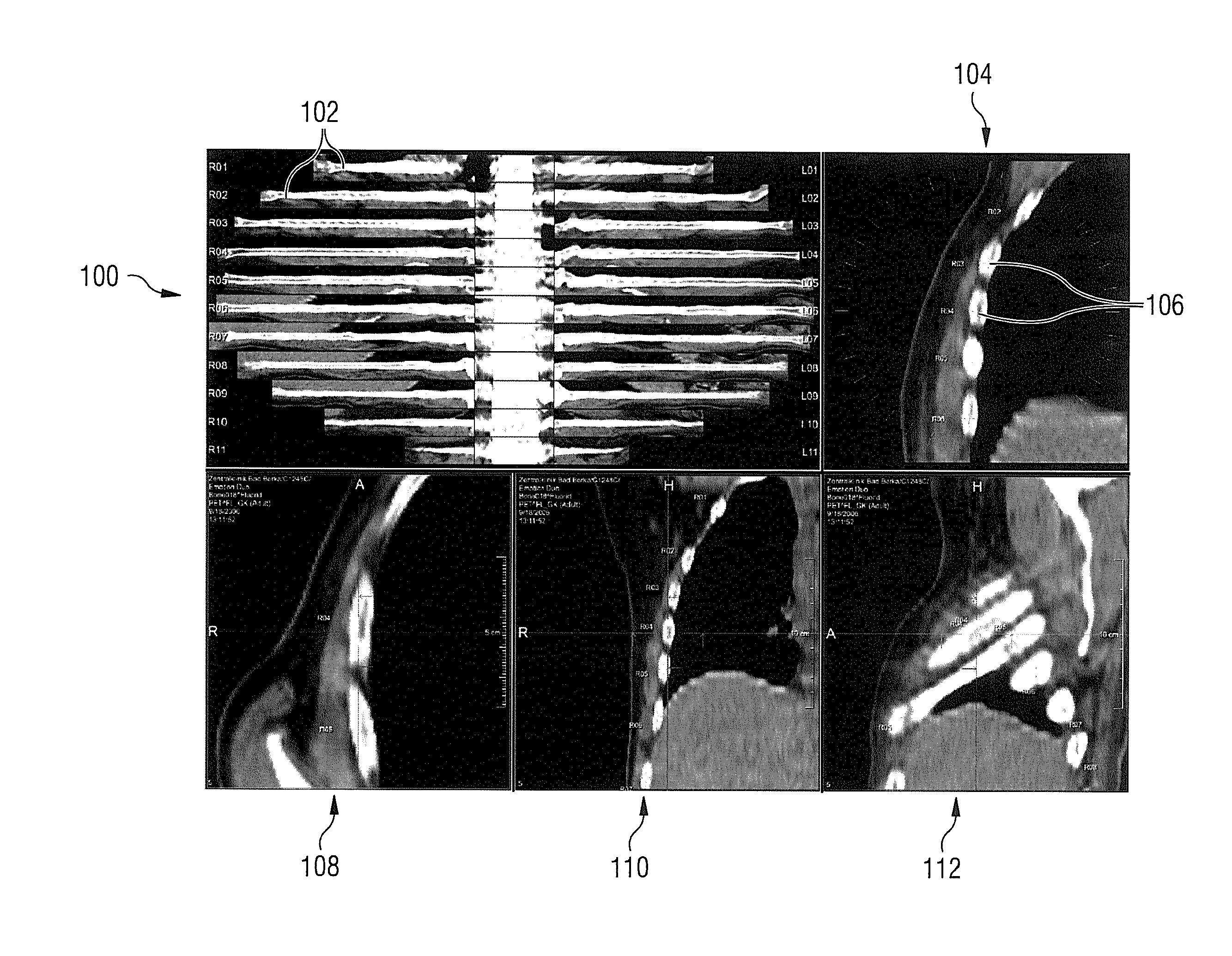
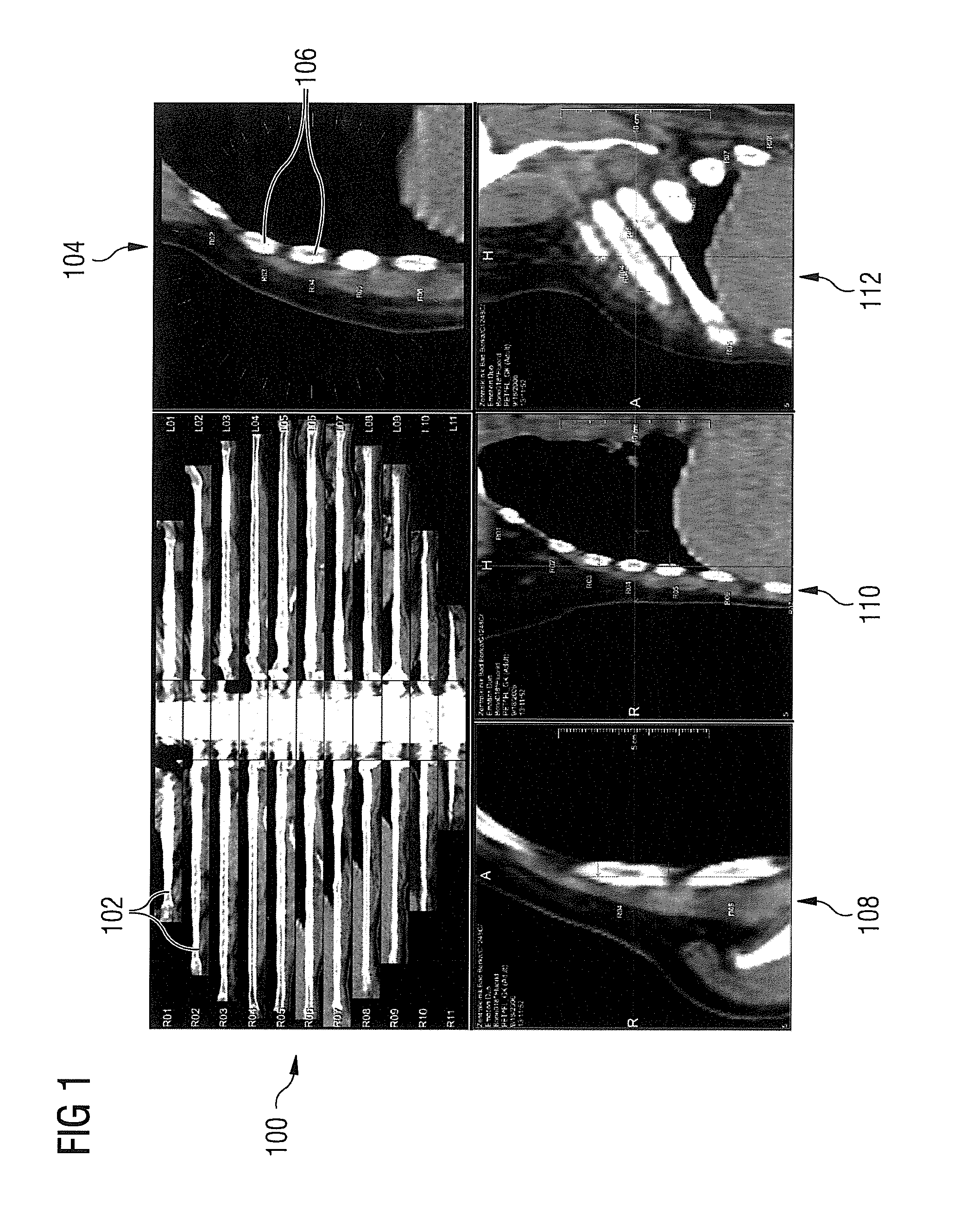
[0056]Embodiments of the invention present methods for enhanced visualizations, automatic / interactive reporting, and follow-up examinations.

[0057]Embodiments of this invention enable the reading physician to easily and efficiently assess which ri...


PUM


Abstract

In a method and apparatus for generating an enhanced image for display from medical imaging data of a subject, a feature of interest in the imaging data elongated in at least one dimension is determined. A location of a line through the imaging data along the feature of interest is obtained, and a projection image along the line is generated from the imaging data.

Description

BACKGROUND OF THE INVENTION[0001]1. Field of the Invention[0002]This invention is directed to a method and apparatus for generating an enhanced image for display from medical imaging data of a subject, in particular for elongated features of interest in an image volume.[0003]2. Description of the Prior Art[0004]The definition of regions or volumes of interest (ROI / VOI) is a typical precursor to quantitative analysis of medical images, such as nuclear medicine emission images (for example, PET or SPECT). Such regions may be defined around areas of high intensity which correspond to high tracer uptake (hotspots). For example, in FDG-PET images for oncology, such areas may be indicative of the presence of a tumor. Oncology physicians frequently annotate lesions in PET scans for the purpose of making a diagnosis, or for use in radiotherapy. The mean or maximum tracer uptake can aid a reader in determining the likelihood of cancer. In longitudinal studies, considering the change in inten...

Claims


Application Information

Patent Timeline
IPC IPC(8): G06T5/00
CPCG06T5/00G06T7/0012G06T2207/30172G06T2207/30008G06T2207/10104G06T7/12G06T7/73G06T2207/30096G06T2207/10108
Inventor KAFTAN, JENSKELLY, MATTHEW DAVIDPLATSCH, GUENTHER
Owner SIEMENS HEALTHCARE GMBH
Why Eureka
Try Eureka