
Method and device for needle visualization

a needle and image technology, applied in the field of medical ultrasound imaging, can solve problems such as delay in needle image display

Active Publication Date: 2013-10-03
GENERAL ELECTRIC CO
View PDF4 Cites 7 Cited by
  • Summary
  • Abstract
  • Description
  • Claims
  • Application Information

AI Technical Summary

Benefits of technology

The present invention provides a method for needle visualization using ultrasound imaging. The method involves acquiring tissue frames alternating with needle frames, and then spatially compounding the frames to create an image frame. To avoid over-enhancing the resulting image, gradual enhancement along the depth of the scan is employed. Additionally, a ratio between values in the needle frame and a new tissue frame is determined and then summed to create a new tissue frame. Overall, this method improves the accuracy and ease of needle visualization during ultrasound imaging.

Problems solved by technology

Although said techniques can present needle images in ultrasound images, there is a delay in the display of needle images.

Method used


Image

  • Method and device for needle visualization
  • Method and device for needle visualization
  • Method and device for needle visualization

Examples


Embodiment Construction

[0018]The present invention will be described in detail by way of specific embodiments. However, it should be understood that the present invention is not limited to these specific embodiments.

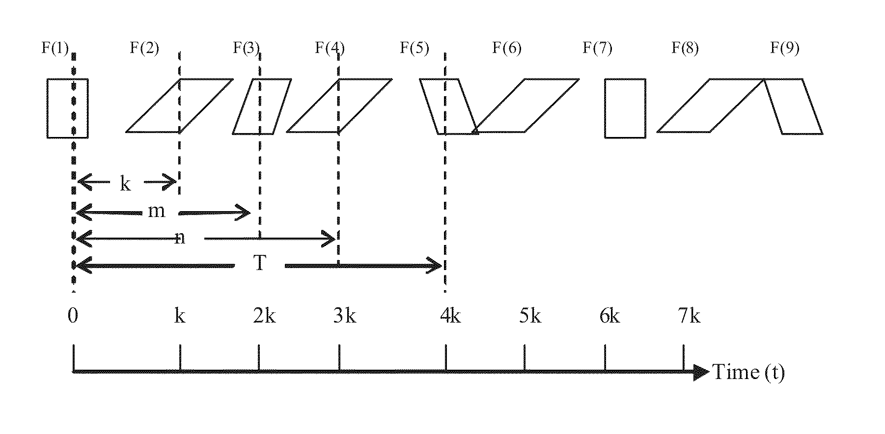
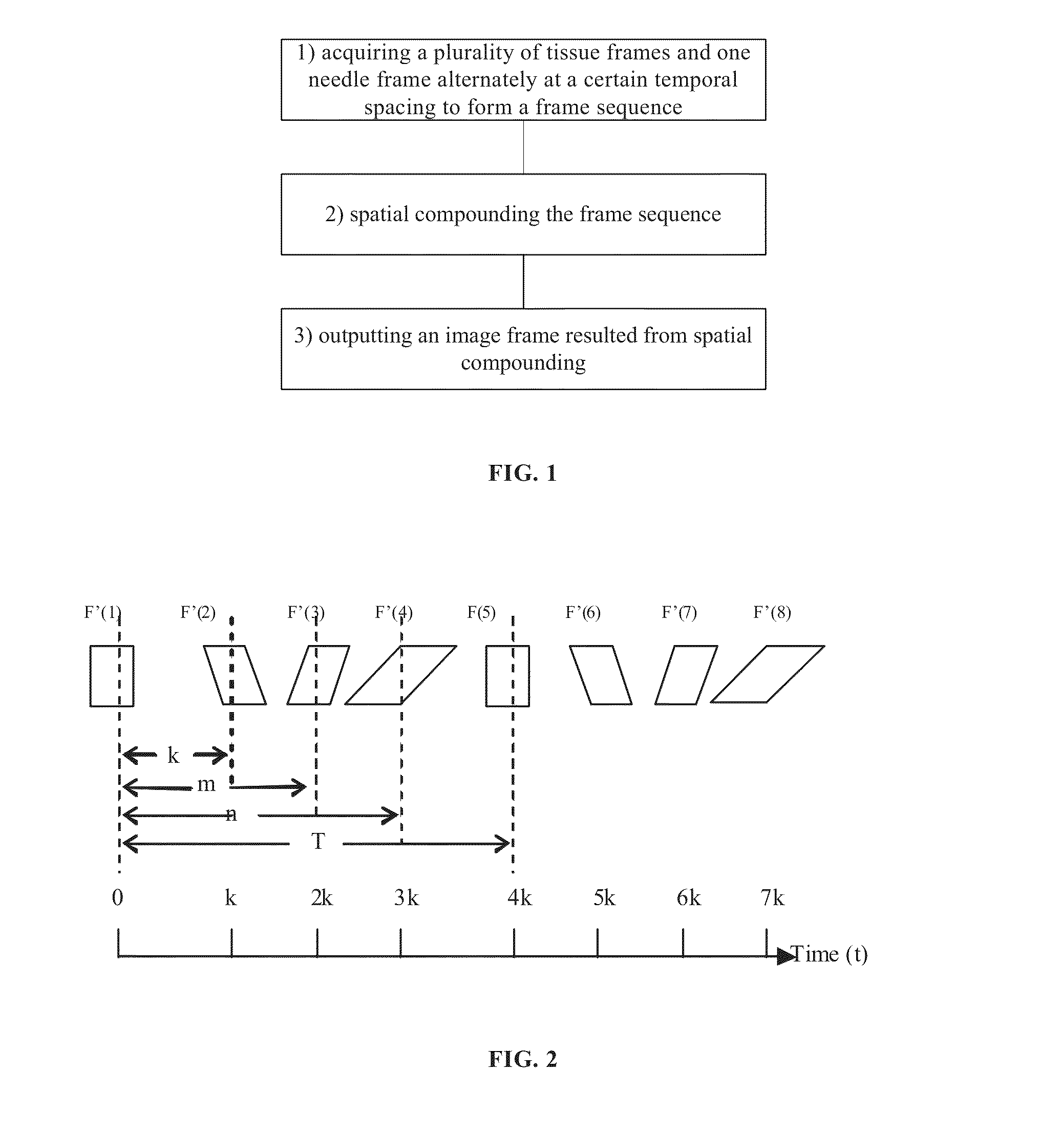
[0019]The prior art technical solution is first introduced here for clarity. As shown in FIG. 1, the prior art technical solution comprises the following steps: (1) acquiring a plurality of tissue frames and one needle frame alternately at a certain temporal spacing to form a frame sequence; (2) spatial compounding the frame sequence; and (3) outputting an image frame resulted from spatial compounding, wherein the plurality generally comprises, for example, three tissue frames obtained through scanning at 0 degree, 15 degrees, and −15 degrees respectively. This is only an example, while scans may be performed at any other angle.

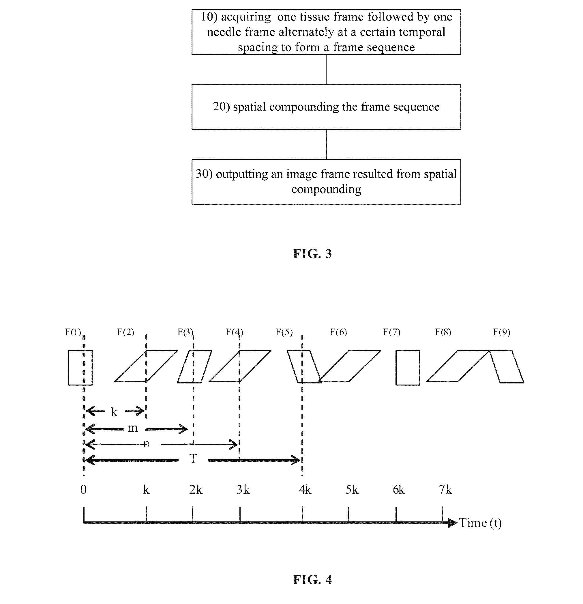
[0020]It should be noted that it is also feasible to acquire four, five, or any other number of, tissue frames followed by one needle frame.

[0021]As shown in FIG. 2, th...


PUM


Abstract

A method for needle visualization for an ultrasound imaging device, the method comprising alternately acquiring a tissue frame followed by a needle frame at a temporal spacing to form a frame sequence, spatially compounding the frame sequence, and outputting an image frame resulting from spatial compounding.

Description

BACKGROUND OF THE INVENTION[0001]1. Field of the Invention[0002]Embodiments of the present invention generally relate to medical ultrasound imaging and, more particularly, to a method and a device for needle visualization.[0003]2. Description of the Prior Art[0004]Since an ultrasound imaging device can provide clear images of human blood vessels and blood flow and is particularly useful as a positioning and aiding tool in procedures such as cardiac invasive therapy, the device enables a physician to discern a lesion site of a patient and each progress of the procedure and is thus crucial to the success of operations.[0005]For procedures demanding invasion into nerves and blood vessels, needle probing and visualization of real-time and high-quality needle images are paramount. Therefore, there exists a need for an ultrasound imaging device to display images of a travelling needle in a real time and high quality fashion.[0006]Existing needle visualization techniques for the ultrasound...

Claims


Application Information

Patent Timeline
Patent Type & Authority Applications(United States)
IPC IPC(8): A61B8/13
CPCA61B8/0841A61B8/5207A61B8/5246A61B8/14A61B8/463G01S15/8995
Inventor GUO, JIANJUNHALMANN, MENACHEMBOWMAN, JONATHAN ALANYANG, JIAJIU
Owner GENERAL ELECTRIC CO
Learn More