
Abnormal syngamy phenotypes observed with time lapse imaging for early identification of embryos with lower development potential

a time-lapse imaging and abnormal syngamy technology, applied in the field of biological and clinical testing, can solve the problems of low birth rate, miscarriage, low birth rate, well-documented adverse outcomes for both mother and fetus, etc., and achieve the effect of lowering the potential for implantation and low development potential

Active Publication Date: 2014-08-07
ARES TRADING SA
View PDF2 Cites 6 Cited by
  • Summary
  • Abstract
  • Description
  • Claims
  • Application Information

AI Technical Summary

Benefits of technology

The patent text describes a method for monitoring embryos to determine their quality during the process of syngamy. By measuring the time between syngamy and the beginning of the first cytokinesis, or the time it takes for the nucleus to divide, researchers can determine which embryos are most likely to make it to the blastocyst stage and implant into the uterus. This method helps to improve the selection of high-quality embryos for implantation and ultimately leads to better outcomes for in vitro fertilization (IVF) procedures.

Problems solved by technology

Infertility is a common health problem that affects 10-15% of couples of reproductive-age.
Multiple gestations have well-documented adverse outcomes for both the mother and fetuses, such as miscarriage, pre-term birth, and low birth rate.
Potential causes for failure of IVF are diverse; however, since the introduction of IVF in 1978, one of the major challenges has been to identify the embryos that are most suitable for transfer and most likely to result in term pregnancy.
The understanding in the art of basic embryo development is limited as studies on human embryo biology remain challenging and often exempt from research funding.
These differences and many others make it inappropriate to directly extrapolate from one species to another.
In spite of such differences, the majority of studies of preimplantation embryo development derive from model organisms and are difficult to relate to human embryo development (Zernicka-Goetz, M.
However, potential risks of these methods also exist in that they prolong the culture period and disrupt embryo integrity (Manipalviratn S, et al.
Not withstanding the recent developments in time lapse imaging that allow clinicians to select embryos with greater developmental potential based on timing parameters of the first few cell cycles, current embryo selection relies primarily on morphological evaluations which are very subjective and offer limited predictive value of embryo viability.
Failure to correctly identify the most viable embryos can lead to unsuccessful IVF treatment or multiple gestation pregnancy.

Method used


Image

  • Abnormal syngamy phenotypes observed with time lapse imaging for early identification of embryos with lower development potential
  • Abnormal syngamy phenotypes observed with time lapse imaging for early identification of embryos with lower development potential
  • Abnormal syngamy phenotypes observed with time lapse imaging for early identification of embryos with lower development potential

Examples


example 1

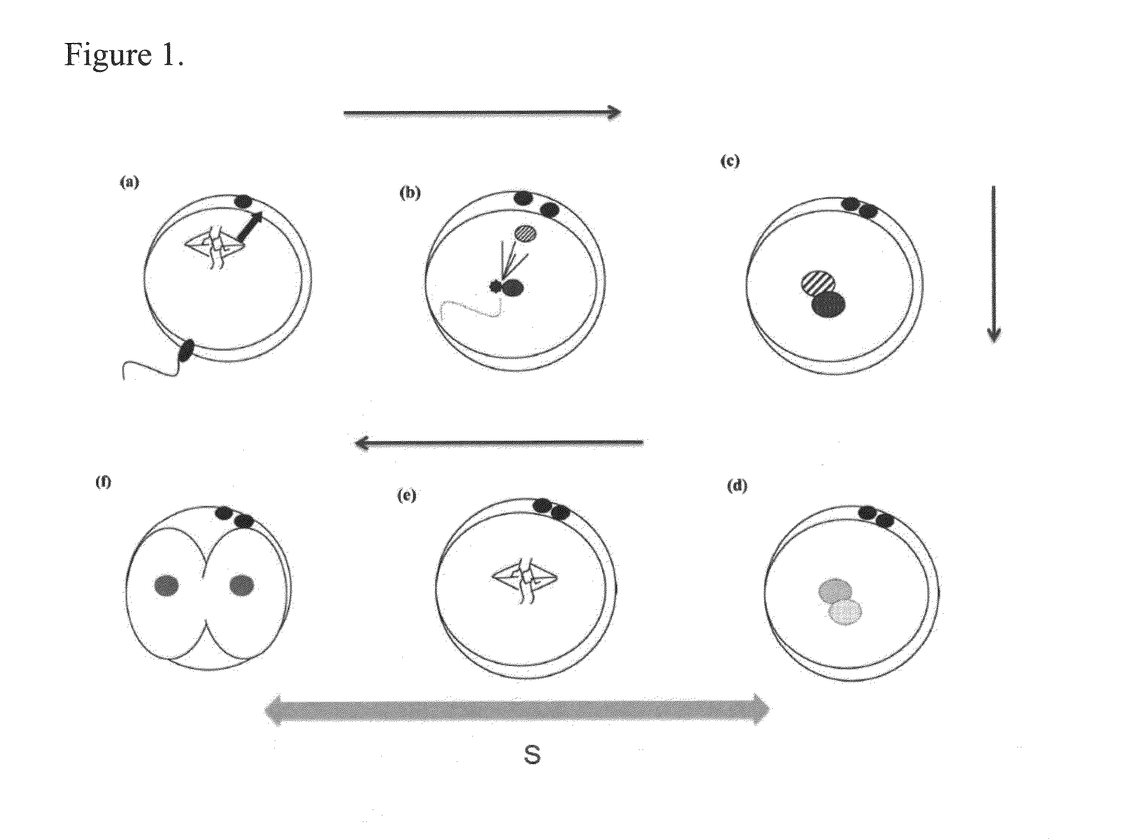
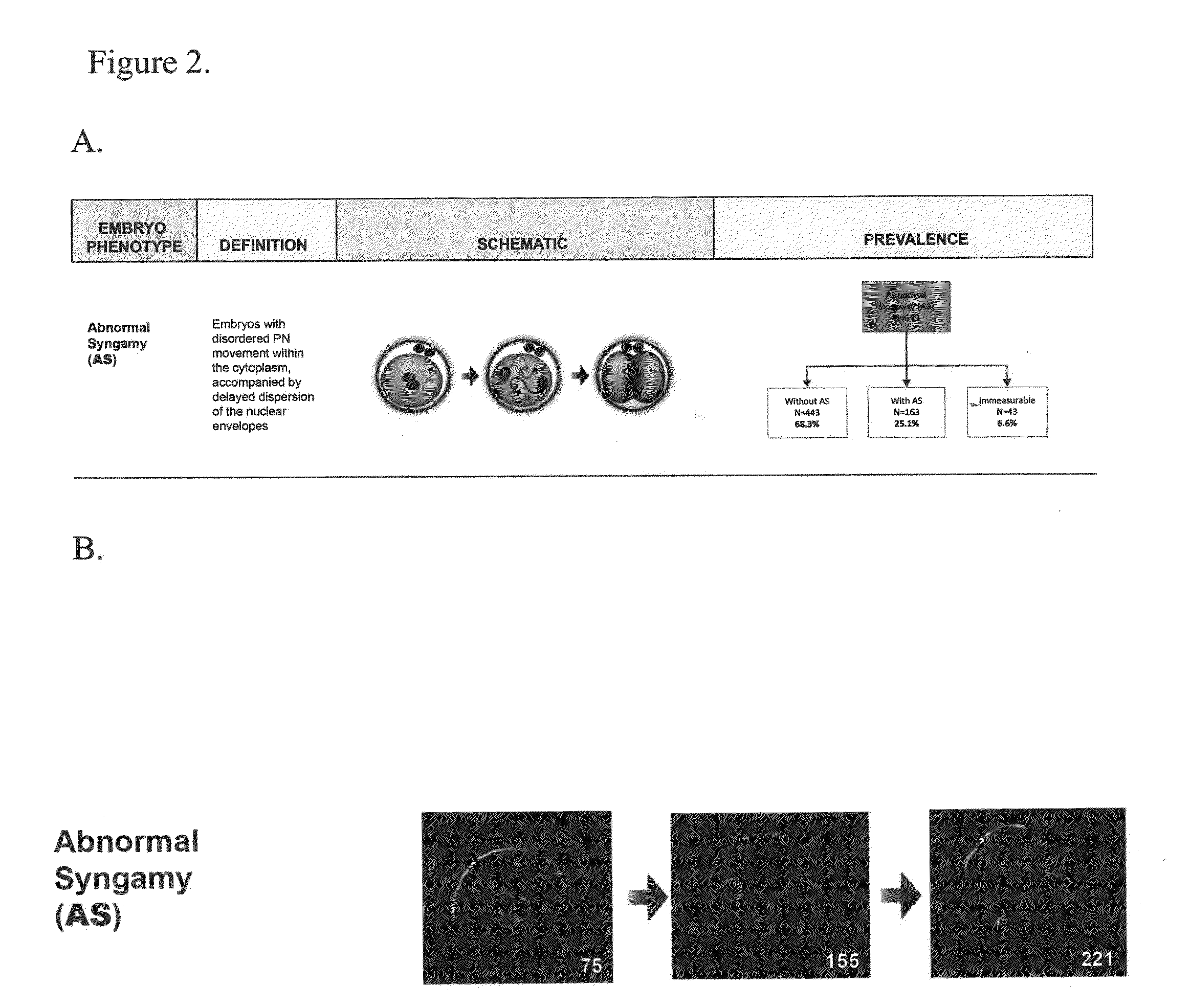
[0107]A retrospective cohort study was done using image data collected from 651 embryos from 67 patients from five clinics in a 16 month period. Patient embryos were imaged using the Eeva™ Test, a time-lapse technology developed for blastocyst prediction.

[0108]All imaged embryos were identified as having 2 pronuclei (PN) before being placed in a multi-well Eeva dish that allows embryos to be tracked individually while sharing a single drop of culture media. A fertilization check was performed according to each clinic's standard protocol. All 2PN embryos were transferred to the Eeva dish immediately after fertilization status was assessed, and the dish was placed on the Eeva scope in the incubator. To maintain a continuous and uninterrupted imaging process from Day 1 through Day 3, no media changes or dish removal from the incubator were permitted. On Day 3, imaging was stopped just before routine embryo grading was performed. All embryos were tracked individually to maintain their i...

example 2

[0115]It has been reported that more than 50% of the human embryos cultured in IVF clinics are aneuploid embryos that have an abnormal number of chromosomes. These aneuploid embryos have lower implantation rates and are not likely to result in live birth of healthy babies. Currently, pre-implantation genetic screening (PGS) is the most effective approach to select against aneuploidy embryos. However, due to its high cost, labor intensive requirements and invasive nature, less than 10% of patients that are treated for assisted reproduction undergo PGS prior to embryo transfer.

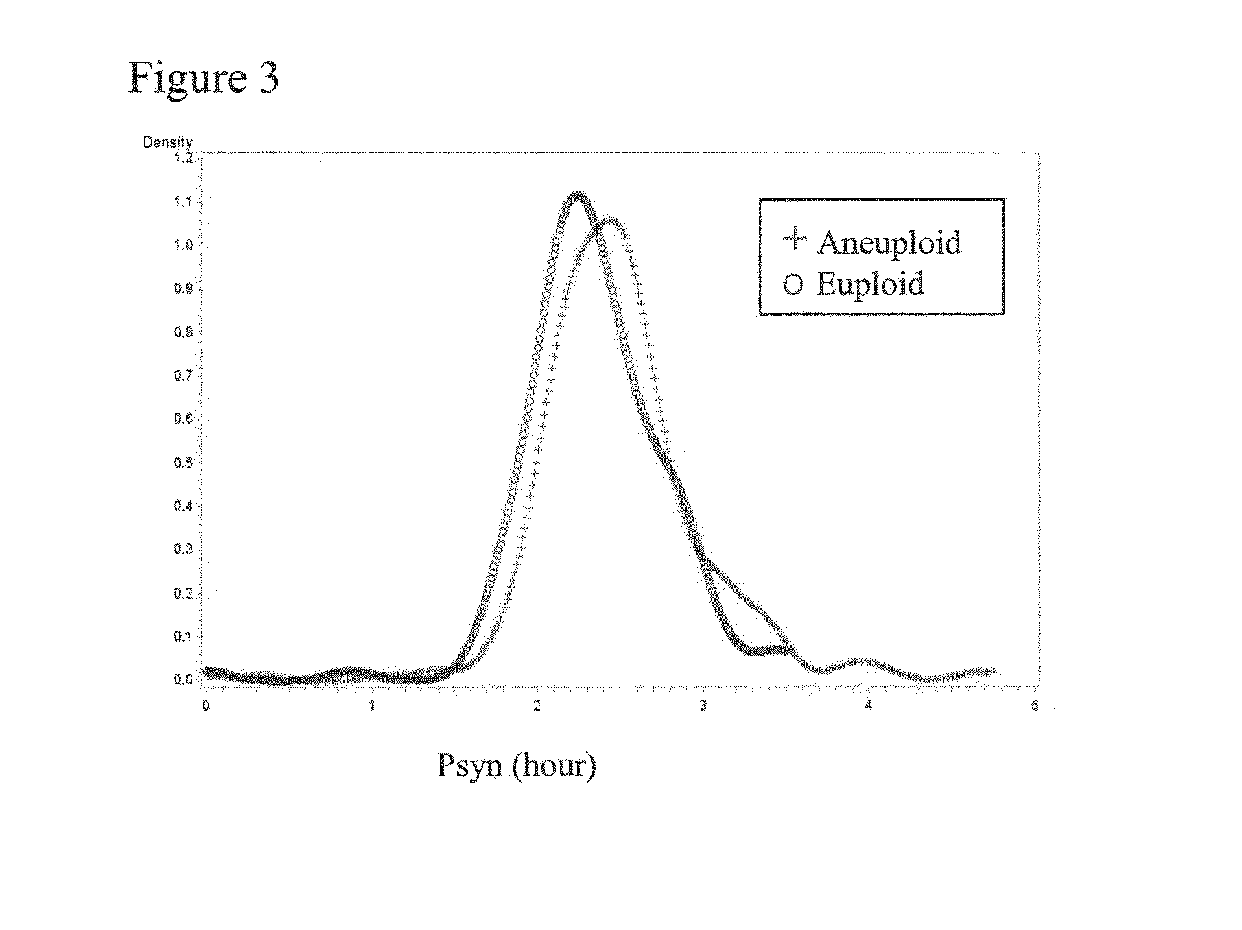
[0116]In order to develop a non-invasive time-lapse enabled assay to assess the risk of embryos being aneuploid, it was discovered that by assessing the Psyn parameter, alone or in combination with other cellular parameter, embryo morphology and patient information, the likelihood of an embryo being aneuploid can be determined.

[0117]By evaluating Psyn values from time-lapse data of 402 embryos, it was discovered...

example 3

[0119]Further analysis of the retrospective cohort study described in the previous examples was performed to evaluate the AS parameters in combination with other atypical phenotype parameters. Using the data from the 67 patients and 639 embryo movies, embryos with one or more atypical phenotypes were analyzed.

[0120]The atypical phenotypes examined were abnormal cleavage (AC), abnormal syngamy (AS), abnormal first cytokinesis (A1cyt), and chaotic cleavage. The AS phenotype was defined as described in Example 1. The AC phenotype was defined as embryos exhibiting producing more than 2 cells during a single cell division event, including AC1 phenotypes that exhibited a first cleavage yielding more than two blastomeres and AC2 phenotypes that exhibited a daughter cell cleavage yielding more than two blastomeres. The A1cyt phenotype was defined as embryos exhibiting oolema ruffling with or without the formation of pseudo furrows before the completion of first cytokinesis. The chaotic clea...


PUM

PropertyMeasurementUnit
timeaaaaaaaaaa
timeaaaaaaaaaa
timeaaaaaaaaaa

Abstract

Methods, compositions and kits for determining the developmental potential of one or more embryos are provided. These methods, compositions and kits find use in identifying embryos in vitro that are most useful in treating infertility in humans.

Description

CROSS REFERENCE TO RELATED APPLICATIONS[0001]This application claims priority to Provisional Appln. No. 61 / 759,598, filed Feb. 1, 2013 and U.S. Provisional Appln. No. 61 / 783,958, filed Mar. 14, 2013, both of which are herein incorporated by reference in their entirety.FIELD OF THE INVENTION[0002]This invention relates to the field of biological and clinical testing, and particularly the imaging and evaluation of zygotes / embryos from both humans and animals.BACKGROUND OF THE INVENTION[0003]Infertility is a common health problem that affects 10-15% of couples of reproductive-age. In the United States alone in the year 2006, approximately 140,000 cycles of in vitro fertilization (IVF) were performed (cdc.gov / art). This resulted in the culture of more than a million embryos annually with variable, and often ill-defined, potential for implantation and development to term. The live birth rate, per cycle, following IVF was just 29%, while on average 30% of live births resulted in multiple ...

Claims


Application Information

Patent Timeline
Patent Type & Authority Applications(United States)
IPC IPC(8): G01N33/50
CPCG01N33/5005G01N2800/367G01N33/5091
Inventor SHEN, SHEHUACHEN KIM, ALICE A.WIRKA, KELLY ATHAYDESURAJ, VAISHALITAN, LEI
Owner ARES TRADING SA
Learn More