Radiation Therapy Guided Using Gamma Imaging

a gamma imaging and guided beam technology, applied in the field of radiotherapy, can solve the problems of collateral damage, anatomical location of tumor movement, and inability to be completely representativ

Inactive Publication Date: 2014-11-20
CUBRESA
View PDF10 Cites 14 Cited by
  • Summary
  • Abstract
  • Description
  • Claims
  • Application Information

AI Technical Summary

Benefits of technology

[0030]It is one object of the invention to provide a method for guiding radiation therapy which can accommodate movement of the lesion caused by respiration and / or cardiac motion and / or peristaltic motion.

Problems solved by technology

Even though MRI provides good location of the tumor at the time of the measurement, these images are normally recorded two to three days prior to the treatment and so may not be completely representative of tumor location on the day of treatment.
This is because the movement of the patient over time can cause the anatomical location of the tumor to move.
This produces collateral damage and may have a significant impact of the quality of life of the patient.
An additional challenge to effective radiation treatment is the effect of motion of the tumor in the body due to respiratory and cardiac motion.
This results in tumor masses moving making the continuous accurate targeting for treatment difficult.
The problem with this is the non-compatibility of linear accelerators and MRI.
This arises because the magnetic field generated by the magnet of course interferes with the operation of the linear accelerator to an extent which cannot be readily overcome.
The problem with this is the non-compatibility of linear accelerators and MRI.

Method used


Image

  • Radiation Therapy Guided Using Gamma Imaging
  • Radiation Therapy Guided Using Gamma Imaging
  • Radiation Therapy Guided Using Gamma Imaging

Examples


Embodiment Construction

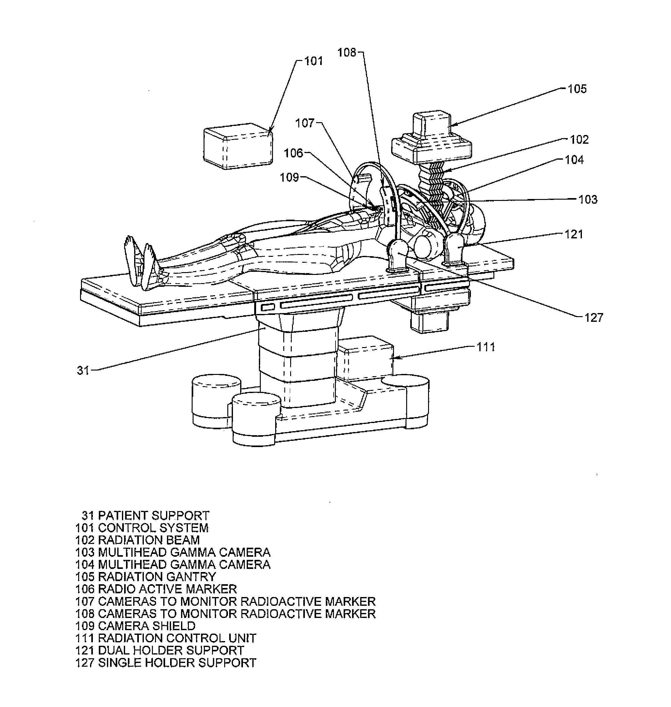
[0089]In FIG. 1 is shown schematically a magnetic resonance imaging system which includes a magnet 10 having a bore 11 into which a patient 12 can be received on a patient table 13. This patient table is in fact a component of the patient support system and this is moved with the patient in the identical position on the patient table as the patient moves from imaging device to treatment device. The system further includes an RF transmit body coil which generates a RF field within the bore. The movable magnet is carried on a rail system with a support suspended on the rail system.

[0090]The system further includes a receive coil system which is located at the isocenter within the bore and receives signals generated from the human body in conventional manner. A RF control system acts to control the transmit body coil and to receive the signals from the receive coil. The two multi-head gamma cameras, 103 and 104, are held in position using camera holders 116 and 117. The same arrangemen...


PUM


Abstract

Radiation therapy of a lesion within a patient is guided to take into account movement of the lesion caused by respiration and/or cardiac effects by using MRI or other imaging system suitable for locating the lesion to image the patient while on the treatment support and using a gamma imaging system responsive to a radiation source preferentially taken up by the lesion and registered with the MRI so as to monitor movement of the lesion in real time and thus guide the beam of the RT.

Description

[0001]This application claims the benefit under 35 USC 119(e) of Provisional Application 61 / 809,648 filed Apr. 8, 2013.[0002]This invention relates to a guided Radiation Therapy system using. GI (Gamma Imaging) in association with another imaging modality such as MRI (Magnetic Resonance Imaging) for location of a lesion for treatment and for controlling the radiation to the lesion.[0003]Reference is made to the following applications of the present applicants, the disclosure of which is incorporated herein by reference[0004]PCT / CA20131050113 filed Feb. 14, 2013[0005]PCTCA2012 / 050423 filed Jun. 26, 2012[0006]U.S. patent application Ser. No. 14 / 128,112 filed on Jun. 26, 2012[0007]PCT / CA2011 / 050074 filed Feb. 10, 2011[0008]Australian Patent Application 2011214864 filed Feb. 10, 2011[0009]Canadian Patent Application 2,788,976 field Feb. 10, 2011[0010]European Patent Application 11741794.9 filed Sep. 4, 2012[0011]U.S. patent application Ser. No. 13 / 516,995 field Oct. 1, 2012BACKGROUND OF...

Claims


Application Information

Patent Timeline
Patent Type & Authority Applications(United States)
IPC IPC(8): A61N5/10A61B6/03
CPCA61N5/107A61B6/037A61N5/1039A61B6/032A61B6/4258A61B6/4291A61B6/4417A61B6/4429A61B6/5247A61B6/5264A61N5/1049A61N2005/1052
Inventor SAUNDERS, JOHNSCHELLENBERG, JAMES
Owner CUBRESA
Why Eureka
Try Eureka