Methods for delineating cellular regions and classifying regions of histopathology and microanatomy

a technology applied in the field of methods for delineating cellular regions and classifying regions of histopathology and microanatomy, can solve the problems of inapplicability of present techniques to a large cohort of histology sections, inability to perform existing techniques, and inability to meet the needs of patients

Active Publication Date: 2015-04-23
RGT UNIV OF CALIFORNIA
View PDF2 Cites 56 Cited by
  • Summary
  • Abstract
  • Description
  • Claims
  • Application Information

AI Technical Summary

Benefits of technology

[0009]In one aspect, embodiments disclosed herein provide methods for delineating cell nuclei and classifying regions of histopathology or microanatomy while remaining invariant to batch effects, comprising: providing a plurality of reference images of histology sections; determining a first set of basis functions from the reference images; classifying histopathology or microanatomy of the histology sections by reference to the first set of basis functions or reference to human engineered features; determining a second set of basis functions for delineating cell nuclei from the reference images; and delineating the nuclear regions of the histology sections based on second set of basis functions.

Problems solved by technology

However, outcome-based analysis requires a large cohort, and the performance of the existing techniques is hindered as a result of large technical and biological variations that are always present in such a cohort.
These techniques suffer from robustness as a result of the batch effect (e.g., technical variations in sample preparation) and biological heterogeneity.
As a result, present techniques are not applicable to a large cohort of histology sections that are collected from different laboratories that do not adhere to an identical protocol.

Method used


Image

  • Methods for delineating cellular regions and classifying regions of histopathology and microanatomy
  • Methods for delineating cellular regions and classifying regions of histopathology and microanatomy
  • Methods for delineating cellular regions and classifying regions of histopathology and microanatomy

Examples


example 1

Characterization of Tissue Histopathology via Predictive Sparse Decomposition and Spatial Pyramid Matching

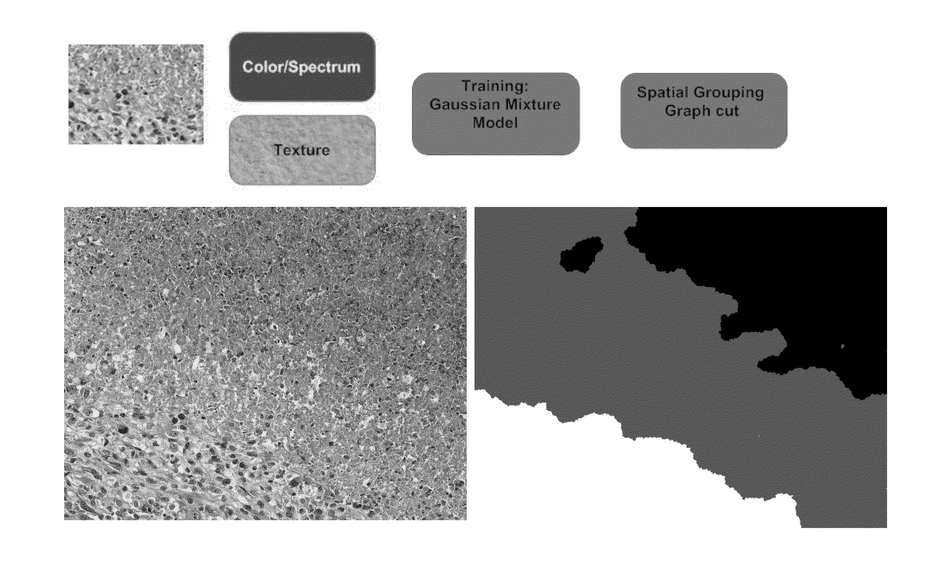
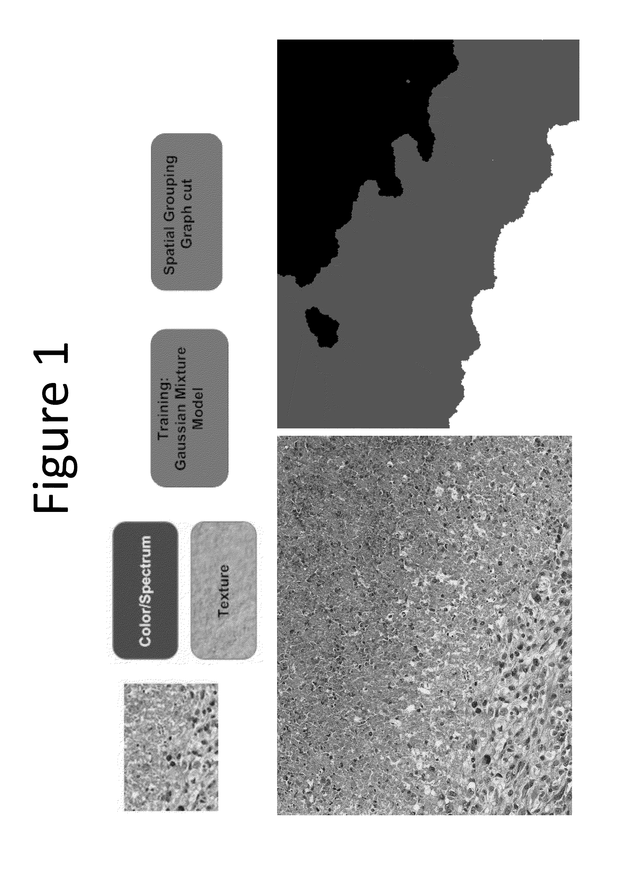
[0038]Disclosed in this example is a tissue classification system and method based on predictive sparse decomposition (PSD) (Kavukcuoglu et al, 2008) and spatial pyramid matching (SPM) (Lazebnik et al, 2006), which utilize sparse tissue morphometric signatures at various locations and scales. Because of the robustness of unsupervised feature learning and the effectiveness of the SPM framework, this method achieved excellent performance even with small number of training samples across independent datasets of tumors. As a result, the composition of tissue histopathology in a whole slide image (WSI) was able to be characterized. In addition, mix grading could also be quantified in terms of tumor composition. Computed compositional indices, from WSI, could then be utilized for outcome based analysis, such as prediction of patient survival statistics or likely responses to therapeut...

example 2

Classification of Tumor Histology Via Morphometric Context

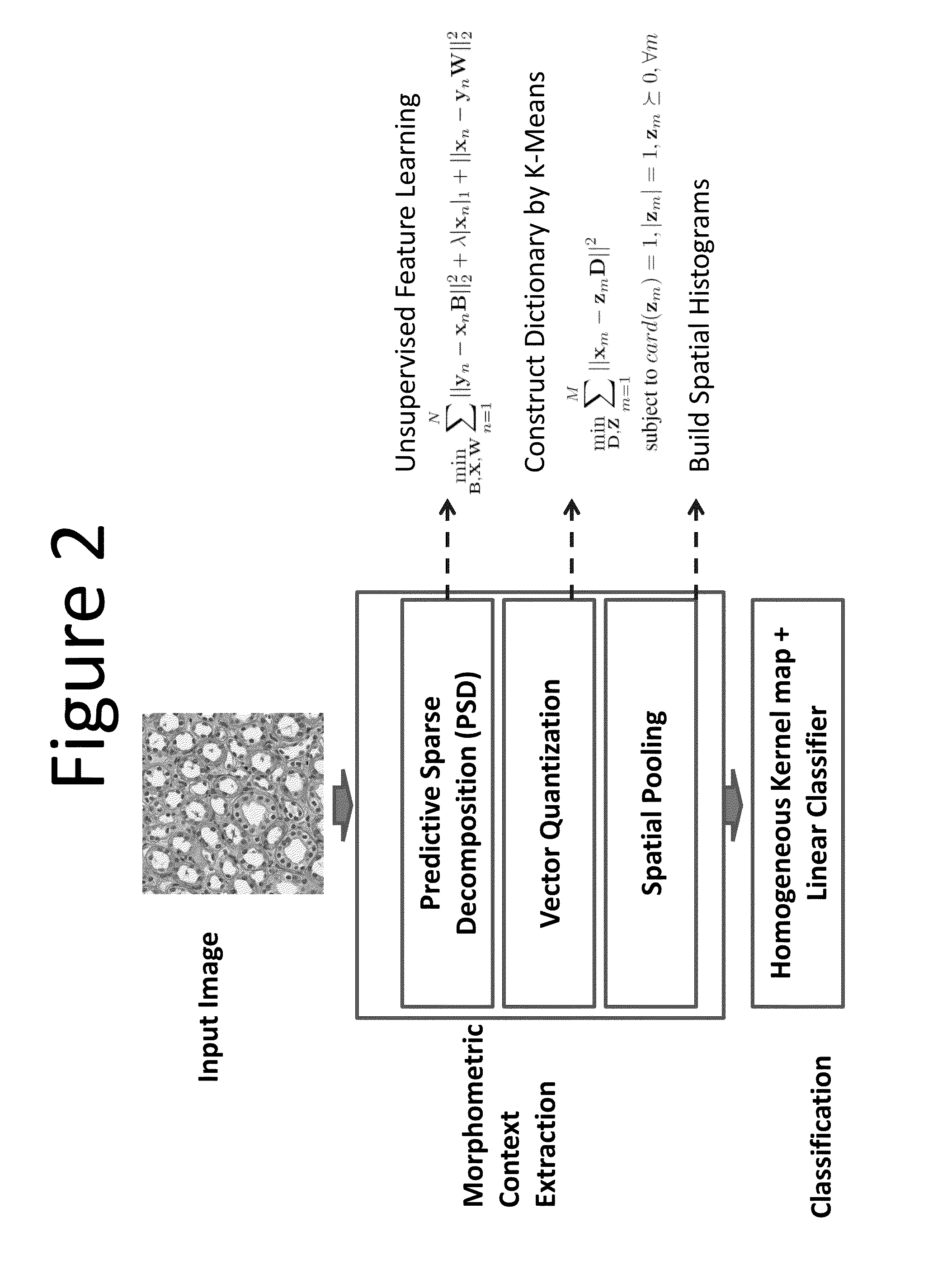
[0073]In this example (Reference: H Chang, A D Borowski, P T Spellman, and B Parvin, “Classification of tumor histopathology via morphometric context,” CVPR 2013), we proposed two variations of tissue classification methods based on representations of morphometric context (one variation is shown in FIG. 2), which were constructed from nuclear morphometric statistics of various locations and scales based on spatial pyramid matching (SPM) (Lazebnik et al, 2006). Due to the effectiveness of our representations, our methods achieved high performance even with a small number of training samples across different segmentation strategies and independent datasets of tumors. The performance was further complemented by the fact that one of the methods had a superior result with linear classifiers. These characteristics dramatically improved the (i) effectiveness of our techniques when applied to a large cohort, and (ii) extensibility to...

example 3

Classification of Histology Sections via Multispectral Convolutional Sparse Coding

[0123]This example (uses a multispectral unsupervised feature learning model (MCSCSPM) for tissue classification, based on convolutional sparse coding (CSC) (Kavukcuoglu et al, 2010) and spatial pyramid matching (SPM) (Lazebnik et al, 2006). The multispectral features are learned in an unsupervised manner through CSC, followed by the summarization through SPM at various scales and locations. Eventually, the image-level tissue representation is fed into linear SVM for efficient classification (Fan et al, 2008). Compared with sparse coding, CSC possesses two merits: 1) invariance to translation; and 2) producing more complex filters, which contribute to more succinct feature representations. Meanwhile, the proposed approach also benefits from: 1) the biomedical intuitions that different color spectrums typically characterize distinct structures; and 2) the utilization of context, provided by SPM, which i...


PUM


Abstract

Embodiments disclosed herein provide methods and systems for delineating cell nuclei and classifying regions of histopathology or microanatomy while remaining invariant to batch effects. These systems and methods can include providing a plurality of reference images of histology sections. A first set of basis functions can then be determined from the reference images. Then, the histopathology or microanatomy of the histology sections can be classified by reference to the first set of basis functions, or reference to human engineered features. A second set of basis functions can then be calculated for delineating cell nuclei from the reference images and delineating the nuclear regions of the histology sections based on the second set of basis functions.

Description

INCORPORATION BY REFERENCE TO ANY PRIORITY APPLICATIONS[0001]This application claims priority to U.S. Provisional Patent Application No. 61 / 880,965 filed on Sep. 22, 2013, and U.S. Provisional Patent Application No. 62 / 017,770 filed on Jun. 26, 2014, both of which are hereby incorporated by reference in their entireties.STATEMENT REGARDING FEDERALLY SPONSORED R&D[0002]This invention was made with government support under Contract No. DE-ACO2-05CH11231 awarded by the U.S. Department of Energy and under Grant Nos. CA1437991 and CA140663 awarded by the National Institutes of Health. The government has certain rights in the invention.BACKGROUND[0003]1. Field of the Invention[0004]The present invention relates to systems and methods for classification of histology composition and delineation of cellular regions while remaining invariant to the batch effects via deep learning and sparse coding.[0005]2. Description of the Related Art[0006]Tissue sections are often stained with hematoxylin ...

Claims


Application Information

Patent Timeline
Patent Type & Authority Applications(United States)
IPC IPC(8): G06K9/00G06K9/62G06V10/772
CPCG06K9/6267G06K9/00147G06V20/698G06V10/7715G06V10/772G06F18/28G06F18/2136
Inventor PARVIN, BAHRAMCHANG, HANGZHOU, YIN
Owner RGT UNIV OF CALIFORNIA
Why Eureka
Try Eureka