
Diagnosis support system, information processing method, and program

a technology of information processing and diagnosis support, applied in the field of diagnosis support apparatus for the brain, can solve problems such as the difficulty of presenting useful information for diagnosis or confirming what abnormality is caused

Inactive Publication Date: 2016-10-20
CANON KK
View PDF5 Cites 10 Cited by
  • Summary
  • Abstract
  • Description
  • Claims
  • Application Information

AI Technical Summary

Benefits of technology

The present invention is related to a diagnosis support system that helps to analyze brain images and provide medical case information. The system utilizes a three-dimensional image of the brain to identify different anatomical regions and searches a database of medical cases to find information about the specific regions. This information is then displayed for diagnosis purposes. Overall, the system can automate the process of analyzing brain images and provide useful information for medical diagnosis.

Problems solved by technology

However, there are various anatomical regions other than the parahippocampal gyrus in the brain, and it is difficult to confirm what abnormality is caused or to present useful information for diagnosis by comprehensively examining each of the anatomical regions.

Method used


Image

  • Diagnosis support system, information processing method, and program
  • Diagnosis support system, information processing method, and program
  • Diagnosis support system, information processing method, and program

Examples


Embodiment Construction

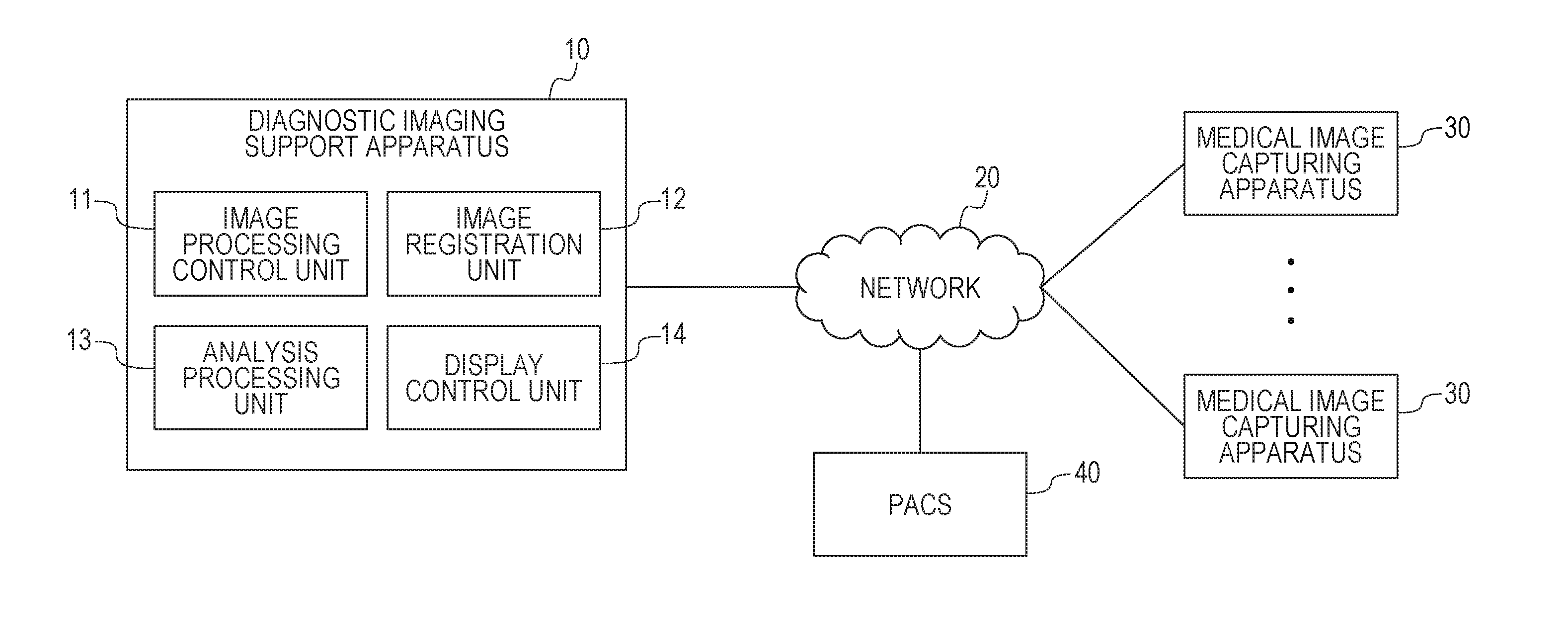
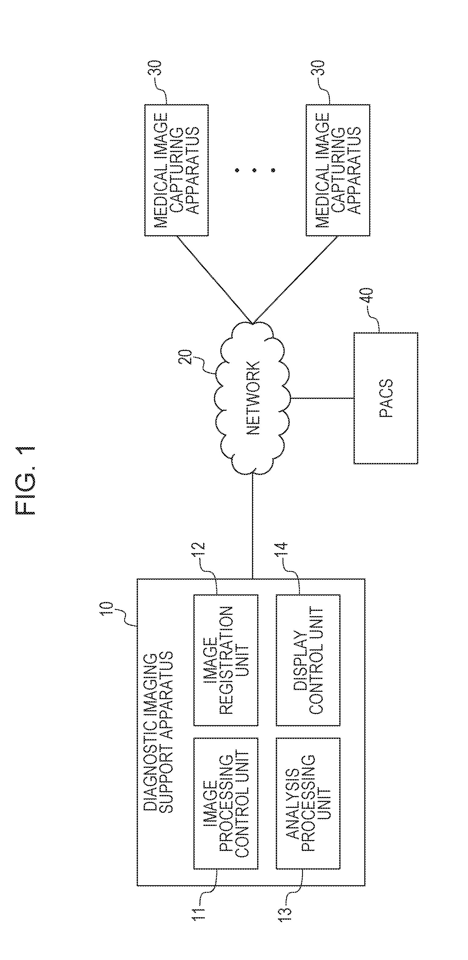
[0021]A diagnostic imaging support system according to one of exemplary embodiments includes a diagnostic imaging support apparatus 10, a network 20, at least one medical image capturing apparatus 30, and a PACS 40. The diagnostic imaging support apparatus 10 has functions including, for example, an image processing apparatus 50, and a display apparatus 60 which will be described below. There is no limitation to such an example, and provided is a concept that diagnostic imaging support apparatus 10 also includes the image processing apparatus 50 itself which does not have the function of the display apparatus 60.

[0022]First, terms necessary for description of the exemplary embodiment of the invention will be described.

[0023]The network 20 in the first embodiment is a line by which respective apparatuses are connected, and examples thereof include a dedicated line, a local area network (LAN), a wireless LAN, and the Internet line.

[0024]The medical image capturing apparatus 30 in the ...


PUM


Abstract

A diagnostic imaging support system that enables confirmation of abnormalities in anatomical regions of a brain includes capturing images of the brain, specifying regions in the captured images that correspond to the anatomical regions, searching a medical database for similar cases based information associated with the specified regions, and displaying the results of the search.

Description

BACKGROUND[0001]1. Field[0002]Aspects of the present disclosure generally relate to a diagnosis support apparatus for a brain, a diagnosis support system, an information processing method, and a program.[0003]2. Description of the Related Art[0004]Processing in which with a use of an image obtained by imaging of an object by an image capturing apparatus such as a CT apparatus or an MRI apparatus, image regions corresponding to anatomical regions are specified in the image, and then information of characteristics of the anatomical regions is obtained has been performed. For example, a technique of measuring atrophy of parahippocampal gyrus from an MRI image obtained by imaging of a brain is disclosed in “Utility of Axial Images in an Early Alzheimer Disease Diagnosis Support System (VSRAD)”, Japanese Journal of Radiological Technology, Vol. 62, No. 9, pp. 1339-1344, 2006.[0005]However, there are various anatomical regions other than the parahippocampal gyrus in the brain, and it is d...

Claims


Application Information

Patent Timeline
IPC IPC(8): G06F19/00G06F17/30G16Z99/00
CPCG06F19/345G06F17/3028G06F19/3443G06F17/30268G06F19/322G06F19/321G16H10/60G16H50/70G16H50/20G16Z99/00G16H30/20
Inventor MIZOBE, HIDEAKI
Owner CANON KK
Learn More