Method and System for Whole Body Bone Removal and Vascular Visualization in Medical Image Data

a technology of image data and vascular visualization, applied in image enhancement, angiography, instruments, etc., can solve the problems of inability to achieve practical use, prohibitive and long operating time required for manual editing, and significant challenges in the automatic segmentation and removal of bone structures from image data

Active Publication Date: 2016-11-10
SIEMENS HEATHCARE GMBH
View PDF22 Cites 71 Cited by
  • Summary
  • Abstract
  • Description
  • Claims
  • Application Information

AI Technical Summary

Problems solved by technology

However, the tedious and long operating time required for manual editing is prohibitive for it to be of practical use.
Furthermore, due to image resolution and noise in the image data, bones and

Method used


Image

  • Method and System for Whole Body Bone Removal and Vascular Visualization in Medical Image Data
  • Method and System for Whole Body Bone Removal and Vascular Visualization in Medical Image Data
  • Method and System for Whole Body Bone Removal and Vascular Visualization in Medical Image Data

Examples


Embodiment Construction

[0023]The present invention relates to a method and system for whole body bone removal and vasculature visualization in medical image data of a patient. Embodiments of the present invention are described herein to give a visual understanding of the bone removal and vasculature visualization method. A digital image is often composed of digital representations of one or more objects (or shapes). The digital representation of an object is often described herein in terms of identifying and manipulating the objects. Such manipulations are virtual manipulations accomplished in the memory or other circuitry / hardware of a computer system. Accordingly, is to be understood that embodiments of the present invention may be performed within a computer system using data stored within the computer system.

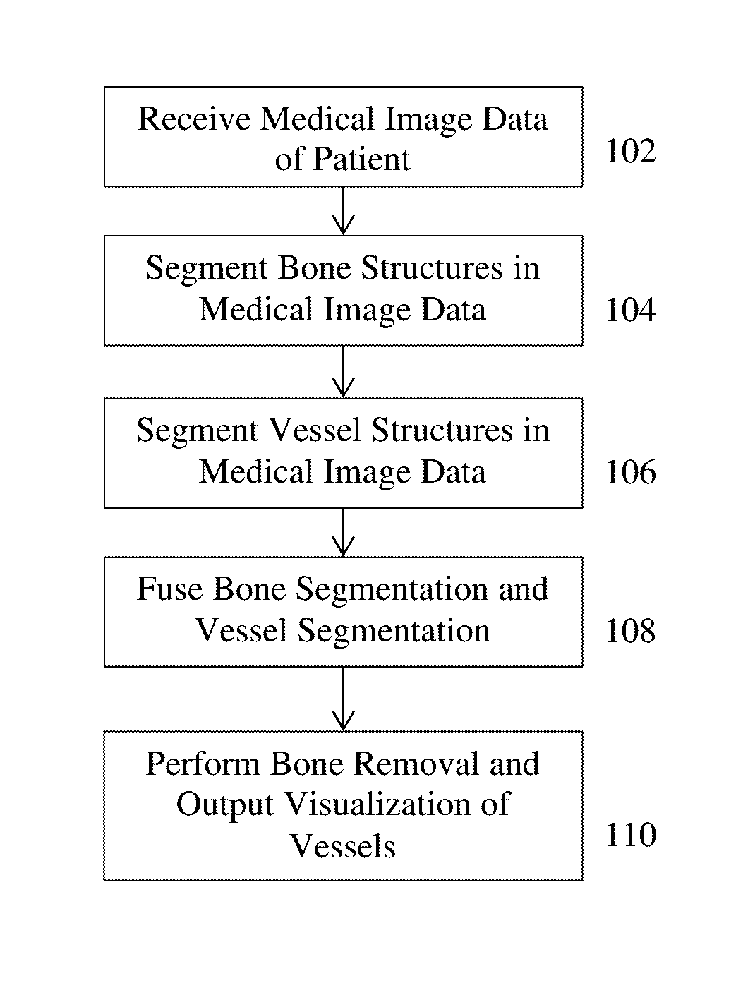
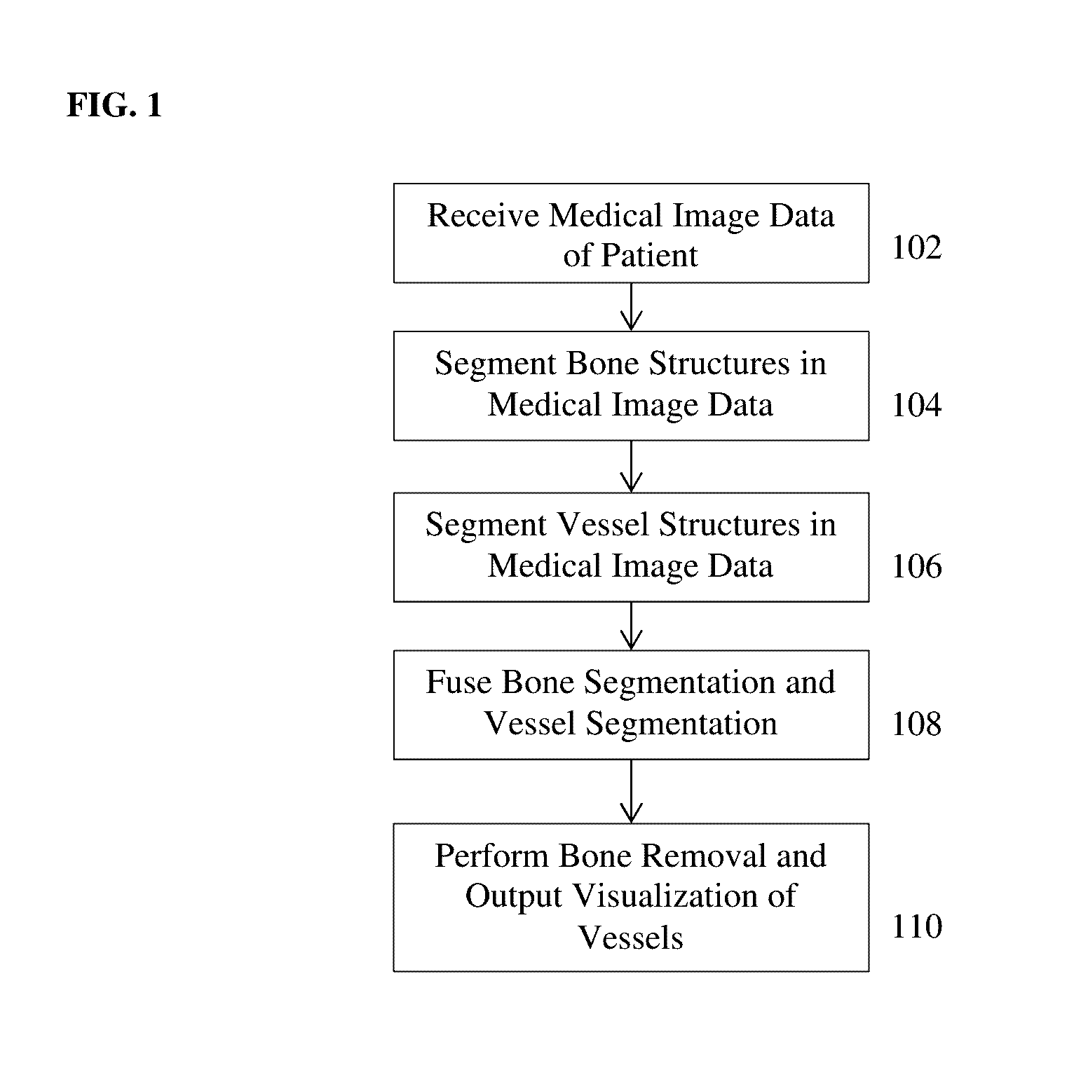
[0024]FIG. 1 illustrates a method for whole body bone removal and vasculature visualization in medical image data according to an embodiment of the present invention. The method of FIG. 1 transfor...


PUM


Abstract

A method and apparatus for whole body bone removal and vasculature visualization in medical image data, such as computed tomography angiography (CTA) scans, is disclosed. Bone structures are segmented in the a 3D medical image, resulting in a bone mask of the 3D medical image. Vessel structures are segmented in the 3D medical image, resulting in a vessel mask of the 3D medical image. The bone mask and the vessel mask are refined by fusing information from the bone mask and the vessel mask. Bone voxels are removed from the 3D medical image using the refined bone mask, in order to generate a visualization of the vessel structures in the 3D medical image.

Description

BACKGROUND OF THE INVENTION[0001]The present invention relates to bone removal and vascular visualization in medical image data, and more particularly, to removal of bone voxels from 3D computed tomography angiography images in order to visualize vessels in the 3D computed tomography angiography images.[0002]Computed Tomography Angiography (CTA) is a medical imaging technique that is often used to visualize the blood vessels in a patient's body. Computed tomography (CT) combines a series of X-ray images taken from different angles and uses computer processing to create cross-sectional images, or slices, of bones, blood vessels and soft tissues inside the body. The cross-sectional images, or slices, can be combined to generate 3D CT volumes. In CTA, a contrast agent is injected into the bloodstream of the patient prior to CT imaging in order to generate contrast enhanced CT images that visualize the patient's vessels.[0003]In CTA images, there is an overlapping intensity between dist...

Claims


Application Information

Patent Timeline
IPC IPC(8): G06T7/00G06K9/62G06K9/46G06K9/66G06T5/50G06T15/08G06V10/50G06V10/764
CPCG06T7/0081G06T5/50G06T15/08G06K9/4604G06T2207/30101G06K9/6267G06T2207/10012G06T2207/10081G06T2207/30008G06K9/66A61B6/5217A61B6/5252A61B6/481A61B6/504G06T5/005G06T2200/04G06T19/00G06T2210/41G06T7/11G06V10/446G06V10/50G06V2201/033G06V2201/03G06V10/764G06V10/7715G06F18/24323G16H30/40
Inventor LAY, NATHANLIU, DAVIDZHOU, SHAOHUA KEVINGEIGER, BERNHARDZHANG, LIORDY, VINCENTXU, DAGUANGSCHWEMMER, CHRISWOLBER, PHILIPPEL-ZEHIRY, NOHA YOUSSRY
Owner SIEMENS HEATHCARE GMBH
Why Eureka
Try Eureka