Anti-tigit antibodies

a technology of anti-tigit antibodies and antibodies, which is applied in the field of anti-tigit antibodies, can solve the problems of poor prognosis, and the overexpression of pd-l1 in tumors, and achieve the effects of stimulating antigen-specific t-cell production, and reducing tumor specific t cell exhaustion

Inactive Publication Date: 2017-07-13
MERCK SHARP & DOHME CORP
View PDF1 Cites 41 Cited by
  • Summary
  • Abstract
  • Description
  • Claims
  • Application Information

AI Technical Summary

Benefits of technology

[0018]In another embodiment, the invention provides an antibody or antigen binding fragment that binds to human TIGIT comprising: (i) a heavy chain variable region CDR1 comprising the amino acid sequence of SEQ ID NO: 57; (ii) a heavy chain variable region CDR2 comprising the amino acid sequence of SEQ ID NO: 58; (iii) a heavy chain variable region CDR3 comprising the amino acid sequence of SEQ ID NO: 59; (iv) a light chain variable region CDR1 comprising the amino acid sequence of SEQ ID NO: 60; (v) a light chain variable region CDR2 comprising the amino acid sequence of SEQ ID NO: 61; and (vi) a light chain variable region CDR3 comprising the amino acid sequence of SEQ ID NO: 62. In one embodiment, the antibody or antigen binding fragment thereof is humanized. In one embodiment, the antibody or antigen binding fragment thereof comprises 1, 2 or 3 amino acid substitutions in the heavy chain CDRs (SEQ ID NOs: 57-59) or in the light chain CDRs (SEQ ID NOs: 60-62), and retains one or more of its functional characteristics. In one embodiment, the antibody or antigen binding fragment thereof optionally has at least one of the following functional characteristics: (i) binds to human TIGIT with a KD value of about 1×10⁻⁹ M to about 1×10-12 M as determined by surface plasmon resonance (e.g., BIACORE) or a similar technique (e.g. KinExa or OCTET), (ii) cross-reacts with cynomolgous and rhesus TIGIT; (iii) blocks binding of human TIGIT to human CD155 and human CD112; (iv) increases T cell activation; (v) stimulates antigen-specific T-cell production of IL-2 and IFNγ; (vi) blocks induction of T cell suppression of activation induced by TIGIT ligation with cognate ligands CD155 and CD112.
[0019]In another embodiment, the invention also provides an antibody or antigen binding fragment thereof that binds to human TIGIT comprising: (i) a heavy chain variable region CDR1 comprising the amino acid sequence of SEQ ID NO: 88; (ii) a heavy chain variable region CDR2 comprising the amino acid sequence of SEQ ID NO: 147; (iii) a heavy chain variable region CDR3 comprising the amino acid sequence of SEQ ID NO: 153; (iv) a light chain variable region CDR1 comprising the amino acid sequence of SEQ ID NO: 91; (v) a light chain variable region CDR2 comprising the amino acid sequence of SEQ ID NO: 148; and (vi) a light chain variable region CDR3 comprising the amino acid sequence of SEQ ID NO: 93. In one embodiment, the antibody or antigen binding fragment thereof comprises: (i) a heavy chain variable region CDR1 comprising the amino acid sequence of SEQ ID NO: 88; (ii) a heavy chain variable region CDR2 comprising the amino acid sequence of SEQ ID NO: 89, 96, 97, 98, 99, 100, 101, 102, 103, 104, 105, 106, 107, 108, 109, 110, 111, 134 or 135; (iii) a heavy chain variable region CDR3 comprising the amino acid sequence of SEQ ID NO: 90, 154, 155, 156, 157, 158, 159, 160, 161, 162, 163, 164, 165, 166 or 167; (iv) a light chain variable region CDR1 comprising the amino acid sequence of SEQ ID NO: 91; (v) a light chain variable region CDR2 comprising the amino acid sequence of SEQ ID NO: 92, 112, 113, 114, 115, 116, 117, 118, 119, 120, 121 or 122; and (vi) a light chain variable region CDR3 comprising the amino acid sequence of SEQ ID NO: 93. In one embodiment, the antibody or antigen binding fragment thereof comprises: (i) a heavy chain variable region CDR1 comprising the amino acid sequence of SEQ ID NO: 88; (ii) a heavy chain variable region CDR2 comprising the amino acid sequence of SEQ ID NO: 89, 134 or 135; (iii) a heavy chain variable region CDR3 comprising the amino acid sequence of SEQ ID NO: 90; (iv) a light chain variable region CDR1 comprising the amino acid sequence of SEQ ID NO: 91; (v) a light chain variable region CDR2 comprising the amino acid sequence of SEQ ID NO: 92; and (vi) a light chain variable region CDR3 comprising the amino acid sequence of SEQ ID NO: 93. In one embodiment, the antibody or antigen binding fragment thereof comprises: (i) a heavy chain variable region CDR1 comprising the amino acid sequence of SEQ ID NO: 88; (ii) a heavy chain variable region CDR2 comprising the amino acid sequence of SEQ ID NO: 134; (iii) a heavy chain variable region CDR3 comprising the amino acid sequence of SEQ ID NO: 90; (iv) a light chain variable region CDR1 comprising the amino acid sequence of SEQ ID NO: 91; (v) a light chain variable region CDR2 comprising the amino acid sequence of SEQ ID NO: 92; and (vi) a light chain variable region CDR3 comprising the amino acid sequence of SEQ ID NO: 93. In one embodiment, the antibody or antigen binding fragment thereof is humanized. In one embodiment, the antibody or antigen binding fragment thereof comprises: (i) a heavy chain variable region CDR1 comprising the amino acid sequence of SEQ ID NO: 88; (ii) a heavy chain variable region CDR2 comprising the amino acid sequence of SEQ ID NO: 134; (iii) a heavy chain variable region CDR3 comprising the amino acid sequence of SEQ ID NO: 154; (iv) a light chain variable region CDR1 comprising the amino acid sequence of SEQ ID NO: 91; (v) a light chain variable region CDR2 comprising the amino acid sequence of SEQ ID NO: 92; and (vi) a light chain variable region CDR3 comprising the amino acid sequence of SEQ ID NO: 93. In one embodiment, the antibody or antigen binding fragment thereof is humanized. In one embodiment, the antibody or antigen binding fragment thereof comprises: (i) a heavy chain variable region CDR1 comprising the amino acid sequence of SEQ ID NO: 88; (ii) a heavy chain variable region CDR2 comprising the amino acid sequence of SEQ ID NO: 134; (iii) a heavy chain variable region CDR3 comprising the amino acid sequence of SEQ ID NO: 155; (iv) a light chain variable region CDR1 comprising the amino acid sequence of SEQ ID NO: 91; (v) a light chain variable region CDR2 comprising the amino acid sequence of SEQ ID NO: 92; and (vi) a light chain variable region CDR3 comprising the amino acid sequence of SEQ ID NO: 93. In one embodiment, the antibody or antigen binding fragment thereof is humanized. In one embodiment, the antibody or antigen binding fragment thereof comprises: (i) a heavy chain variable region CDR1 comprising the amino acid sequence of SEQ ID NO: 88; (ii) a heavy chain variable region CDR2 comprising the amino acid sequence of SEQ ID NO: 134; (iii) a heavy chain variable region CDR3 comprising the amino acid sequence of SEQ ID NO: 156; (iv) a light chain variable region CDR1 comprising the amino acid sequence of SEQ ID NO: 91; (v) a light chain variable region CDR2 comprising the amino acid sequence of SEQ ID NO: 92; and (vi) a light chain variable region CDR3 comprising the amino acid sequence of SEQ ID NO: 93. In one embodiment, the antibody or antigen binding fragment thereof is humanized. In one embodiment, the antibody or antigen binding fragment thereof comprises: (i) a heavy chain variable region CDR1 comprising the amino acid sequence of SEQ ID NO: 88; (ii) a heavy chain variable region CDR2 comprising the amino acid sequence of SEQ ID NO: 134; (iii) a heavy chain variable region CDR3 comprising the amino acid sequence of SEQ ID NO: 157; (iv) a light chain variable region CDR1 comprising the amino acid sequence of SEQ ID NO: 91; (v) a light chain variable region CDR2 comprising the amino acid sequence of SEQ ID NO: 92; and (vi) a light chain variable region CDR3 comprising the amino acid sequence of SEQ ID NO: 93. In one embodiment, the antibody or antigen binding fragment thereof is humanized. In one embodiment, the antibody or antigen binding fragment thereof comprises: (i) a heavy chain variable region CDR1 comprising the amino acid sequence of SEQ ID NO: 88; (ii) a heavy chain variable region CDR2 comprising the amino acid sequence of SEQ ID NO: 134; (iii) a heavy chain variable region CDR3 comprising the amino acid sequence of SEQ ID NO: 158; (iv) a light chain variable region CDR1 comprising the amino acid sequence of SEQ ID NO: 91; (v) a light chain variable region CDR2 comprising the amino acid sequence of SEQ ID NO: 92; and (vi) a light chain variable region CDR3 comprising the amino acid sequence of SEQ ID NO: 93. In one embodiment, the antibody or antigen binding fragment thereof is humanized. In one embodiment, the antibody or antigen binding fragment thereof comprises: (i) a heavy chain variable region CDR1 comprising the amino acid sequence of SEQ ID NO: 88; (ii) a heavy chain variable region CDR2 comprising the amino acid sequence of SEQ ID NO: 134; (iii) a heavy chain variable region CDR3 comprising the amino acid sequence of SEQ ID NO: 159; (iv) a light chain variable region CDR1 comprising the amino acid sequence of SEQ ID NO: 91; (v) a light chain variable region CDR2 comprising the amino acid sequence of SEQ ID NO: 92; and (vi) a light chain variable region CDR3 comprising the amino acid sequence of SEQ ID NO: 93. In one embodiment, the antibody or antigen binding fragment thereof is humanized. In one embodiment, the antibody or antigen binding fragment thereof comprises: (i) a heavy chain variable region CDR1 comprising the amino acid sequence of SEQ ID NO: 88; (ii) a heavy chain variable region CDR2 comprising the amino acid sequence of SEQ ID NO: 134; (iii) a heavy chain variable region CDR3 comprising the amino acid sequence of SEQ ID NO: 160; (iv) a light chain variable region CDR1 comprising the amino acid sequen

Problems solved by technology

Notably, overexpression of PD-L1 in tumors is associated w

Method used


Image

  • Anti-tigit antibodies
  • Anti-tigit antibodies
  • Anti-tigit antibodies

Examples


example 1

Generation of Rat and Mouse Anti-hTIGIT Antibodies

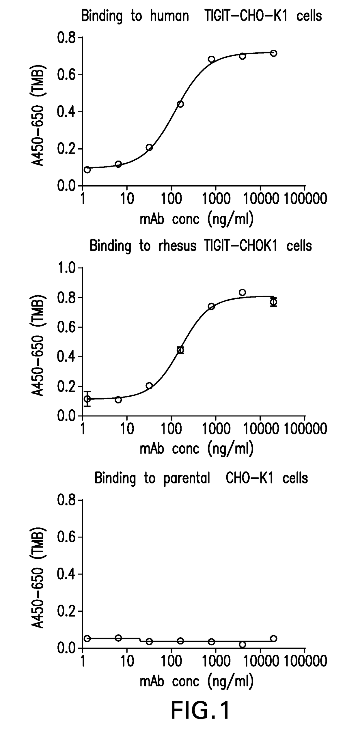
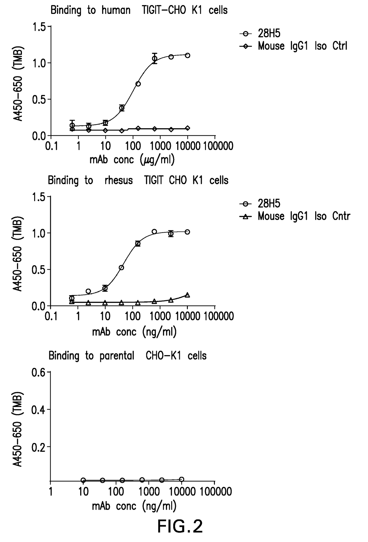
[0370]To generate antibodies to human TIGIT, Lewis rats were immunized with his-tagged human TIGIT recombinant protein from Sino Biologicals Cat#10917-H08H) using RIBI adjuvant and footpad injection on a biweekly schedule. Alternatively, Balb / C mice were immunized with human Fc-tagged human TIGIT recombinant protein using RIBI adjuvant and footpad injection on a biweekly schedule. Immunized animals were bled and serum titers determined for binding to human TIGIT transfected CHOK1 cells using a cell-based ELISA (described below). The animals with the highest titers were given a final boost with recombinant protein and draining popliteal lymph nodes isolated four days later. Hybridomas were generated by electrofusion of isolated lymphocytes with the myeloma fusion partner P3X63-AG8.653 using the Cytopulse Hybrimmune electrofusion system. Fused cells were plated in 96-well plates in DMEM / F12, 15% BCS, HAT, IL-6, OPI supplement, and gent...

example 2

Characterization of Anti-hTIGIT Antibodies

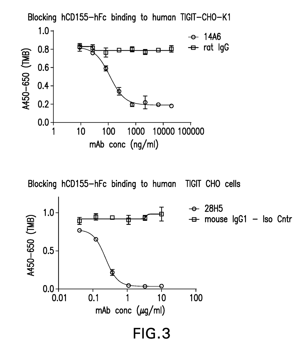
[0374]Supernatants from positive clones were tested for their ability to block recombinant human CD155-huFc protein binding to hTIGIT CHOK1 cells in a cell-based ELISA format. Human TIGIT-CHO-K1 cells were plated in 96-well plates as described above. Media was removed from the plates and 50 μl of hybridoma supernatant was incubated with the human TIGIT CHO-K1 cells at 4° C. for 30 minutes. Fifty microliters of human CD155-huFc was added to the plate for a final concentration of 0.5 μg / ml of human CD155-huFc and incubated for 30 minutes at 4° C. Assay plates were washed 3 times with PBS / 0.05% Tween-20 as above. Binding of human CD155-huFc to the hTIGIT-CHOK1 cells was detected using an HRP-conjugate F(ab)′2 goat anti-human IgG secondary antibody (Jackson 109-036-098) at 1:2000 dilution in CHO-K1 media. Plates were developed using TMB and stopped using TMB Stop Solution as described above and the A450-620 nm determined.

[0375]A rat antibody gen...

example 3

In Vitro T-Cell Activity Assay for Antagonistic Anti-hTIGIT Antibodies

[0381]Of the antibodies tested, approximately 352 monoclonal antibodies shown to block binding of CD155-Fc to CHO cells expressing hTIGIT were screened for their capacity enhance T cell activity in vitro using cell-based functional assays.

[0382]One assay we developed to characterize the functional consequence of blocking human TIGIT receptor utilized Jurkat cells, an immortalized line of human T lymphocyte cells (clone, E6-1; ATCC TIB-152) engineered to over-express human TIGIT (hTIGIT-Jurkat) which were co-cultured with THP-1 cells, a human monocytic cell line in the presence or absence of one of the TIGIT ligands, CD155 and CD112. hTIGIT-Jurkat cells co-cultured with THP-1 cells and stimulated with plate-bound anti-CD3 mAb produce IL-2, but when TIGIT ligand (CD155 or CD112) is added to the co-culture, IL-2 levels were reduced in a ligand-dependent manner. Treatment with antibodies that blocked the CD155- or CD1...


PUM

PropertyMeasurementUnit
Fractionaaaaaaaaaa
Fractionaaaaaaaaaa
Fractionaaaaaaaaaa

Abstract

The present invention relates to anti-TIGIT antibodies, as well as use of these antibodies in the treatment of diseases such as cancer and infectious disease.

Description

FIELD OF THE INVENTION[0001]The present invention relates to anti-TIGIT antibodies, as well as use of these antibodies in the treatment of diseases such as cancer and infectious disease.BACKGROUND OF THE INVENTION[0002]A key factor for enabling tumor immunotherapy emerged from discoveries that inhibitory immune modulatory receptors (IMRs), that generally function as immune checkpoints to maintain self-tolerance, are central to the ability of tumor microenvironments to evade immunity. Blockade of inhibitory IMRs appears to unleash potent tumor-specific immune responses more effectively than direct stimulation of tumor-immunity with activating cytokines or tumor vaccines, and this approach has the potential to transform human cancer therapy. An important implication and opportunity now arises for the potential to develop new antibody antagonists for other IMRs and to combine antagonist antibodies to more than one IMR in order to increase the proportion of responders in oncology clinic...

Claims


Application Information

Patent Timeline
IPC IPC(8): C07K16/28A61K45/06A61K39/395
CPCC07K16/2803C07K16/2818A61K39/3955A61K45/06C07K2317/565A61K2039/507C07K2317/92C07K2317/24C07K2317/52C07K2317/76C07K2317/56C07K16/28A61K2039/505C07K2317/34C07K2317/21C07K2317/74A61P31/00A61P35/00A61P37/02A61P37/04A61P43/00Y02A50/30C07K2317/73
Inventor WILLIAMS, SYBIL M. G.LAFACE, DRAKEFAYADAT-DILMAN, LAURENCERAGHUNATHAN, GOPALANLIANG, LINDASEGHEZZI, WOLFGANG
Owner MERCK SHARP & DOHME CORP
Why Eureka
Try Eureka