Systems and methods for meso-dissection

a meso-dissection and slide technology, applied in image enhancement, image data processing, instruments, etc., can solve the problems of manual labor in the process of generating these annotations, and the system lacks the flexibility to extract tissue from multiple tissue slides

Active Publication Date: 2017-11-09
ROCHE MOLECULAR SYST INC
View PDF0 Cites 12 Cited by
  • Summary
  • Abstract
  • Description
  • Claims
  • Application Information

AI Technical Summary

Benefits of technology

[0008]Therefore, the proposed workflow makes it possible for all the labeled tissue slides from tissue blocks or patients being digitized on a high-resolution digital pathology system in a batch assembly mode and saved to an image management system. And for a pathologist or a medical professional to use the digital pathology software application offline on any computer to import, review the digital images and annotate on the images of the chosen reference slides and select the set of reference annotations for each of the slides to be milled. Once the reference annotations are outlined, inter-marker registration operation is executed on the computer to batch execute on the all tissue datasets and output the corresponding milling annotations on the images of the slides to be milled and save them to an image management system. In an immediate or later session, a meso-dissection system technician or operator, without any further guidance or input from the medical professional, can physically load all the labelled milling slides and load them on the meso-dissection system and connect to the image management system to import the milling images and associated annotations. And for each of the slide to be physically milled, using the software interface on the dissection system, use same-marker registration operation to map milling annotations to the camera coordinate system of the live capture and collect the extracted tissue into the labelled container. Therefore, this overall workflow makes the digital pathology-guided meso-dissection system operational in a high-volume pre-clinical and clinical setting.

Problems solved by technology

Existing systems are designed to be operated in a sequential manner one slide at a time by a skilled professional, and the process of generating these annotations is a manually intensive process.
These systems lack the flexibility to extract tissue from multiple tissue slides in a correlated manner, analyze and investigate molecular and genomic sequence analysis information from multiple regions and adjacent slides is of use of in pre-clinical drug discovery, development investigative studies and clinical prognostic and predictive analysis.

Method used


Image

  • Systems and methods for meso-dissection
  • Systems and methods for meso-dissection
  • Systems and methods for meso-dissection

Examples


embodiment 1

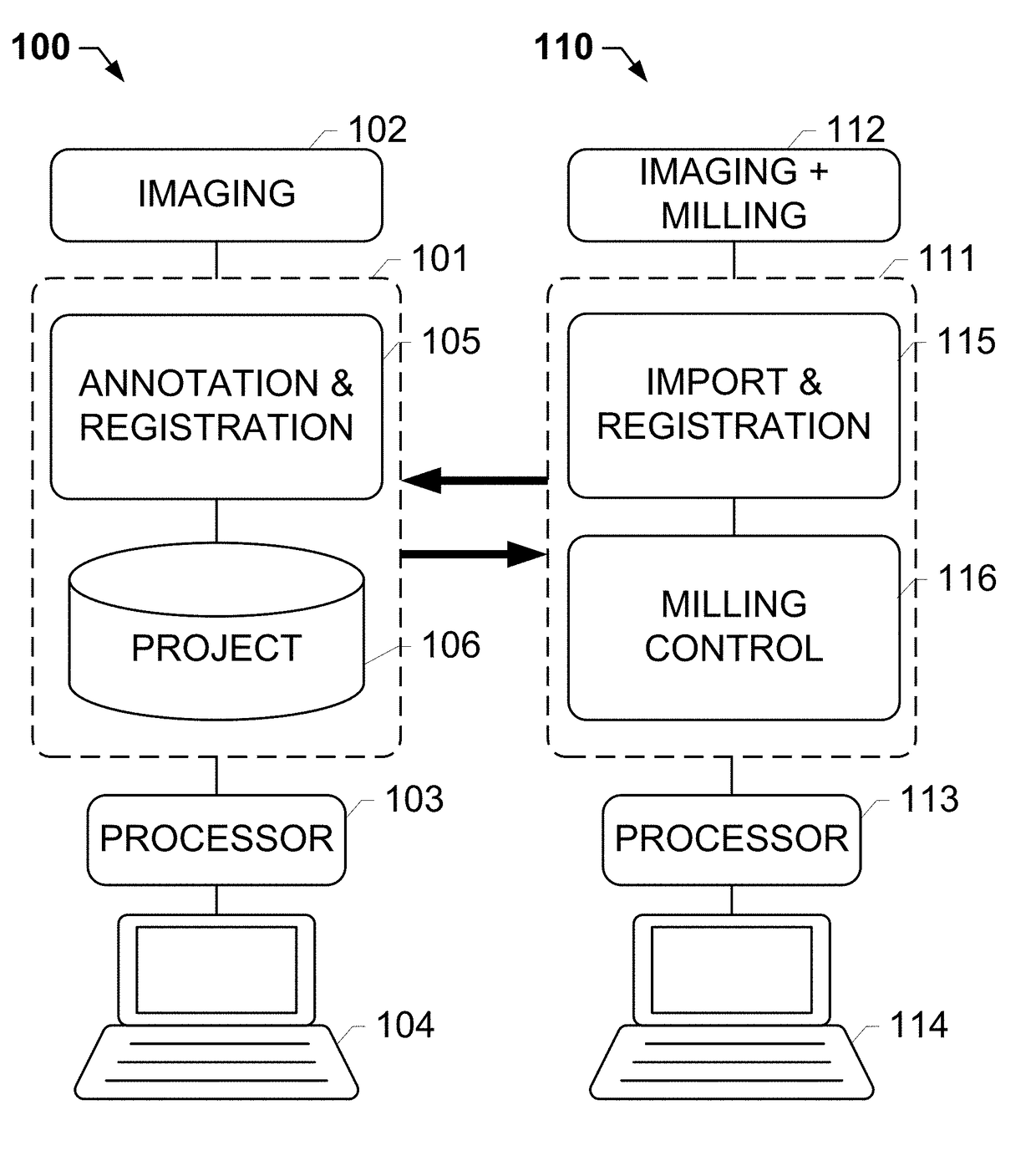
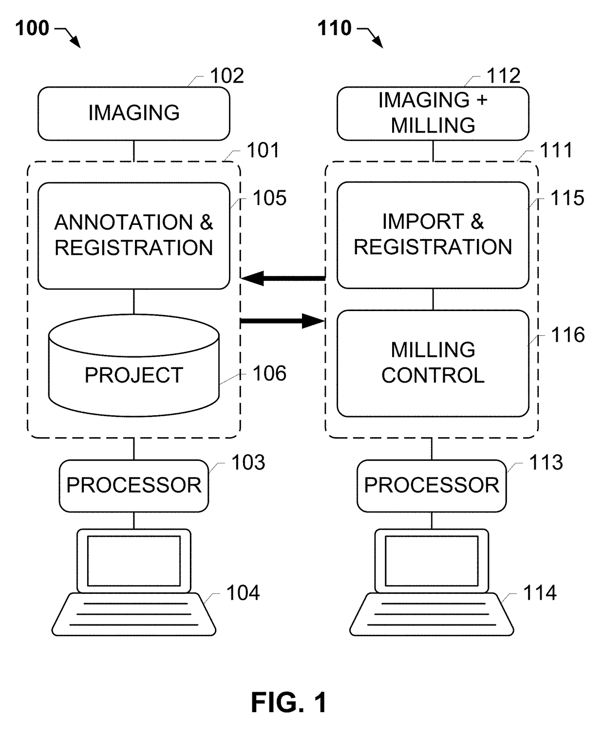
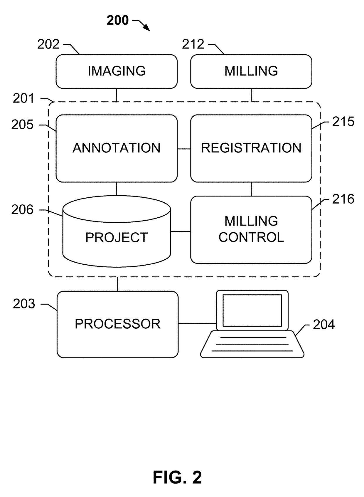
[0076]An instrument for combined digital pathology and meso-dissection, comprising: a processor; and a memory coupled to the processor, the memory to store computer-readable instructions that, when executed by the processor, cause the processor to perform operations comprising: using an intermarker registration operation to map annotations from a first image to one or more of a plurality of adjacent images including a milling image; and subsequently dissecting a milling slide corresponding to the milling image based on the annotations; wherein the annotations are mapped to a live-image of the milling slide. The instrument of embodiment 1, wherein the first image and the plurality of adjacent images correspond to serial sections of a tissue block.

[0077]The instrument of any one of the preceding embodiments, wherein annotated areas of interest on the milling slide are subsequently dissected based on the annotated live image.

[0078]The instrument of any one of the preceding embodiments,...

embodiment 7

[0082]The instrument of embodiment 7, wherein the associating comprises using a unique identifier.

[0083]The instrument of embodiment 7 or 8, wherein a result of analysis of the milled tissue sample is also associated with the patient.

[0084]The instrument of any one of the preceding embodiments, wherein a result of analysis of the milled tissue sample is used to determine one or more of a subsequent scanning operation or a subsequent milling operation.

[0085]A digital pathology instrument, comprising: a processor; and a memory coupled to the processor, the memory to store computer-readable instructions that, when executed by the processor, cause the processor to perform operations comprising: annotating a first image of a plurality of serial images of a tissue block; mapping one or more annotations from the first image to a milling image; and exporting the one or more annotations of the milling image to a milling system.

embodiment 11

[0086]The instrument of embodiment 11, wherein the milling system dissects a milling slide associated with the milling image based on the one or more annotations and / or wherein the milling system maps the annotations from the annotated milling image to a live image of a milling slide using a same-marker registration operation.

[0087]The instrument of embodiment 11 or 12, wherein the mapping of annotations from the first image to the milling image uses an intermarker registration operation.

[0088]A meso-dissection instrument, comprising: a processor; and a memory coupled to the processor, the memory to store computer-readable instructions that, when executed by the processor, cause the processor to perform operations comprising: importing one or more annotations from an imaging system, wherein the one or more milling annotations indicate regions of interest of a tissue specimen, said tissue specimen being one of a plurality of serial sections of a tissue block, and wherein said one or ...


PUM

PropertyMeasurementUnit
areasaaaaaaaaaa
flexibilityaaaaaaaaaa
digital pathology imagingaaaaaaaaaa

Abstract

The subject disclosure provides systems, computer-implemented methods, and clinical workflows for meso-dissection of biological specimens and tissue slides by incorporating annotation and inter-marker registration modules within digital pathology imaging and meso-dissection (or milling) systems. Images of a reference slide a milling slide may be acquired using the same imaging system, with the annotations on the image associated with the milling slide being based on the inter-marker registration. Each image along with its respective annotations and meta-data may be associated with a project or a case, and stored in an image management system. A same-marker registration may be used to map annotations from the annotated image of the milling slide to a live image of the milling slide. The milling slide may be milled based on the annotations, with milled tissue output into a contained that is labeled in association with the labeled input slides.

Description

CROSS-REFERENCE TO RELATED APPLICATIONS[0001]This patent application is a continuation of International Patent Application No. PCT / EP2016 / 051894 filed Jan. 29, 2016, which claims priority to and the benefit of U.S. Provisional Application No. 62 / 110,477 filed Jan. 31, 2015. Each of the above patent applications are hereby incorporated by reference herein in their entireties.FIELD OF THE SUBJECT DISCLOSURE[0002]The present subject disclosure relates to imaging for medical diagnosis. More particularly, the present subject disclosure relates to automated micro-dissection (meso-dissection) of slide-mounted tissue slides guided by digital pathology.BACKGROUND OF THE SUBJECT DISCLOSURE[0003]In the analysis of biological specimens such as tissue sections, blood, cell cultures and the like, biological specimens are mounted on a slide, stained with one or more combinations of stain and biomarkers, and the resulting assay is viewed or imaged for further analysis. Observing the assay enables a...

Claims


Application Information

Patent Timeline
Patent Type & Authority Applications(United States)
IPC IPC(8): G01N1/28G06T7/00G06T7/33G01N1/38
CPCG01N1/286G06T7/0014G06T7/33G06T2207/30024G01N2001/383G06T2207/10056G01N2001/2873G01N2800/00
Inventor BARNES, MICHAELCHUKKA, SRINIVASQADRI, MOHAMMAD
Owner ROCHE MOLECULAR SYST INC
Why Eureka
Try Eureka