

Antibody molecules for cancer treatment
a technology for anti-cancer and antibody molecules, applied in the field of new anti-cancer treatment antibodies, can solve the problems of large patient response to treatment, suffer from existing anti-pd1 antibody molecules, etc., and achieve the effects of improving the activation of t cells, prolonging the terminal half-life, and surprising and advantageous properties
- Summary
- Abstract
- Description
- Claims
- Application Information
AI Technical Summary
Benefits of technology
Problems solved by technology
Method used
Image

Examples
example 1
n of Mouse Antibodies Binding to PD1 and Blocking PDL-1 Binding to PD1
[0311]The extracellular domain (ECD) of human PD1 protein (amino acids 21-170 of GenBank accession number AAO63583.1) was synthesized and prepared as an immunogen. Mice were then immunized and then boosted with human PD1 ECD protein following standard laboratory immunization techniques.
[0312]Plasma was then harvested from the immunized mice and screened to identify individuals with sufficient titre of anti-PD1 immunoglobulin. Following standard laboratory methods, lymphocytes from the selected mice were harvested and fused with mouse myeloma cells to generate hybridomas. These hybridomas were subsequently screened to identify hybridomal lines which produced antibody molecules which exhibited binding in low nM affinity range to human PD1 (ECD), and also blocked the binding of human PD-L1 to PD1, using the assays described herein below.
[0313]Several hybridomas which produced antibodies that bound to human PD1 and bl...
example 2
n of Humanized Anti-PD1 Antibodies
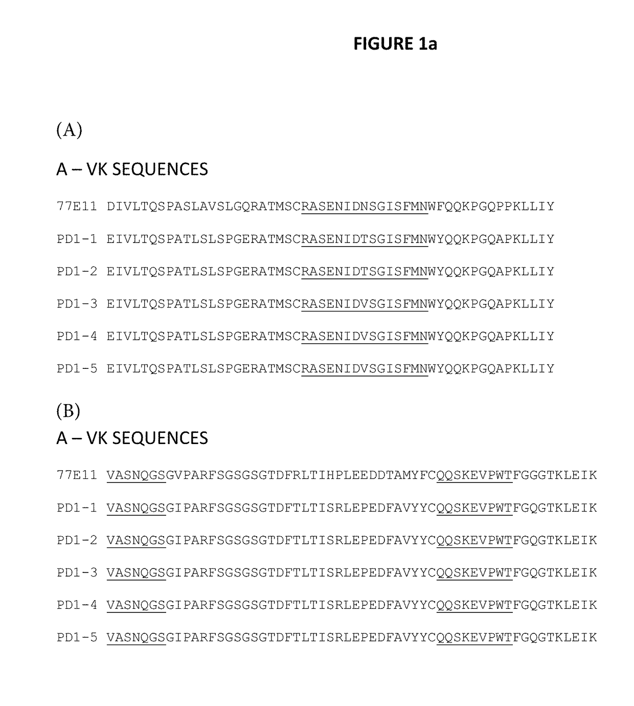
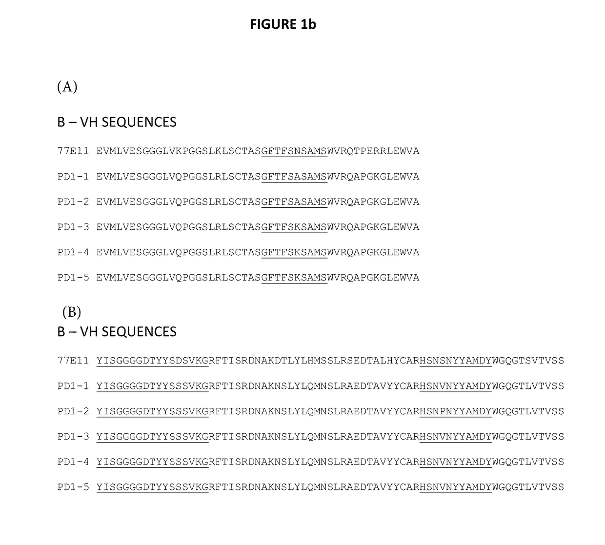
[0316]V-genes of murine 77E11 hybridomal cell line were identified following PCR and sequencing protocols well known in the art. The V-genes were then fused to closest matching human germline genes, using standard molecular biology techniques. In the case of 77E11, this resulted in chimeric murine / human antibody molecules having of mouse Vk and Vh amino acid residues fused to human Ck and Ch1 amino acid residues.
[0317]Once prepared, the chimeric murine / human antibody was then subjected to “humanization” protocols. Here, mutated amino acid libraries of the chimeric 77E11 Fab clones were prepared. Additional library positions were also prepared so as to remove sequence liabilities (i.e. amino acid residues known to be immunogenic or to create potential manufacturing problems). These mutated libraries usually have 5 to 10 binary (mouse vs human) positions per V-region. Several smaller libraries can be built to address specific liabilities in more compl...
example 3
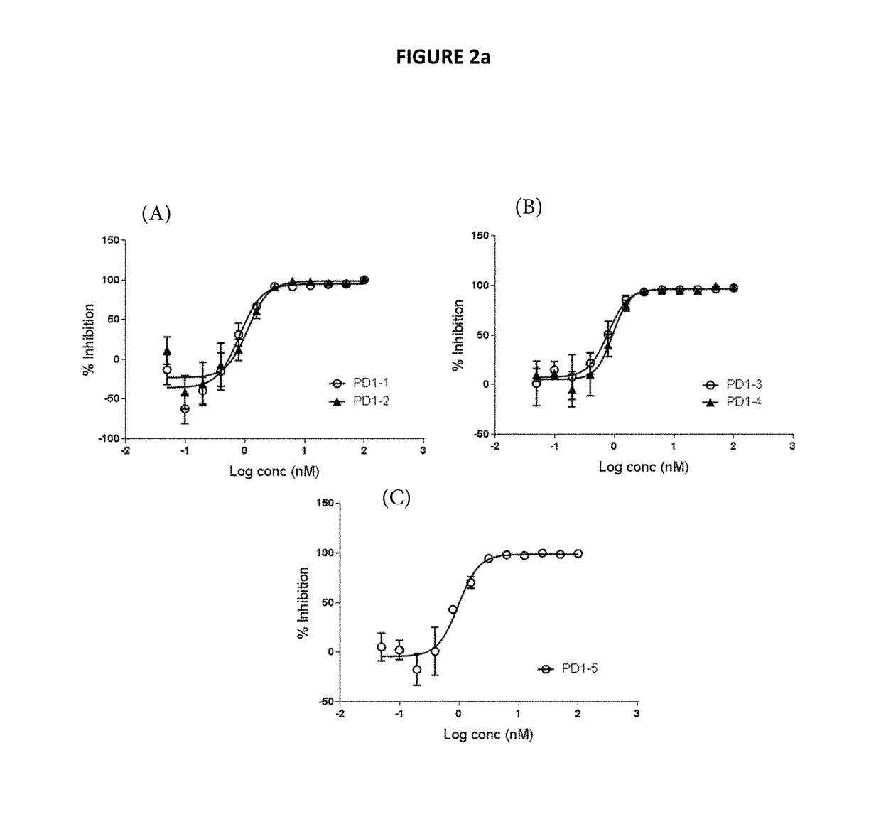
Binding to Human PD1 and Blocking of PD1 Ligand Interaction
[0322]The binding of the representative humanized anti-PD1 antibodies derived from murine 77E11 hybridoma (as described in Example 2) to human PD1 was determined.
[0323]Binding affinities to recombinant monomeric human PD1 were measured by SPR using a ProteOn XPR36 instrument. PD1-1, PD1-2 and PD1-3 antibodies were captured on Protein A / G surfaces that was amine coupled to the surface. Human PD1 ECD was injected over the captured antibodies for 600 sec at a flowrate of 30 ul / min and a dissociation for 1200 sec. The concentrations of human PD1 ECD were 0 nM, 6.25 nM, 12.5 nM, 25 nM, 50 nM, and 100 nM. The background was subtracted from the raw data and sensorgrams were then fit globally to 1:1 Langmuir binding to provide affinity (KD) values.
[0324]Using that protocol, it was determined that antibodies PD1-1, PD1-2 and PD1-3 bind to human PD1 as presented in the table below.
TABLE 2Binding to recombinant human PD1 (KD, nM)Antibo...
PUM
| Property | Measurement | Unit |
|---|---|---|
| Fraction | aaaaa | aaaaa |
| Molar density | aaaaa | aaaaa |
| Molar density | aaaaa | aaaaa |
Abstract
Description
Claims
Application Information