Systems and methods for area-of-interest detection using slide thumbnail images

a technology of area-of-interest and slide thumbnails, applied in image data processing, image enhancement, instruments, etc., can solve the problems of high difficulty in solving the aoi problem, accurate and precise aoi detection, etc., and achieve the effect of efficient and accurate computation of aoi regions

Active Publication Date: 2018-01-11
VENTANA MEDICAL SYST INC
View PDF7 Cites 21 Cited by
  • Summary
  • Abstract
  • Description
  • Claims
  • Application Information

AI Technical Summary

Benefits of technology

[0005]The subject disclosure solves the above-identified problems by providing systems and computer-implemented methods for accurate determination of AOIs for various different types of input slides. Slide thumbnail images (or thumbnails) may be assigned into one of five different types, and separate algorithms for AOI detection may be executed depending on the slide type, with the goal being to execute operations that can efficiently and accurately compute AOI regions from whole slide thumbnail images, since a single generic solution is unable to account for all the data variability. Each thumbnail may be divided into one of 5 types based on the tissue morphology, layout, organization of tissue on the slide thumbnail, etc. The slide types include ThinPrep (RTM) slides, also referred to as “cell line slides”, having a single disc structure (or any other similarly stained slide), tissue micro-array (TMA) slides having a grid structure, control slides having faint tissue (for a controlHER2 tissue slide, there exists a specified number of circular cores, usually 4), smear slides having tissue diffused throughout the slide, and a default or generic slide. A control slide is a slide type comprising both a tissue sample (that shall actually be examined and analyzed) and a control tissue sample that is typically used for verifying histological techniques and reagent reactivity. It comprises a plurality of tissue regions (cores) of the tissue sample to be actually analyzed at defined positions along a straight line.

Problems solved by technology

Unfortunately, the first step in existing methods of acquiring a thumbnail image prior to any further analysis is limited to using low resolution thumbnail cameras, and accurate and precise AOI detection is highly challenging, particularly given the variability of data with different types of slides having different stain intensities, morphology, tissue organization, etc.
The use of a single AOI detection methodology for a wide variety of slides has made solving of the AOI problem very difficult.

Method used


Image

  • Systems and methods for area-of-interest detection using slide thumbnail images
  • Systems and methods for area-of-interest detection using slide thumbnail images
  • Systems and methods for area-of-interest detection using slide thumbnail images

Examples


embodiment 1

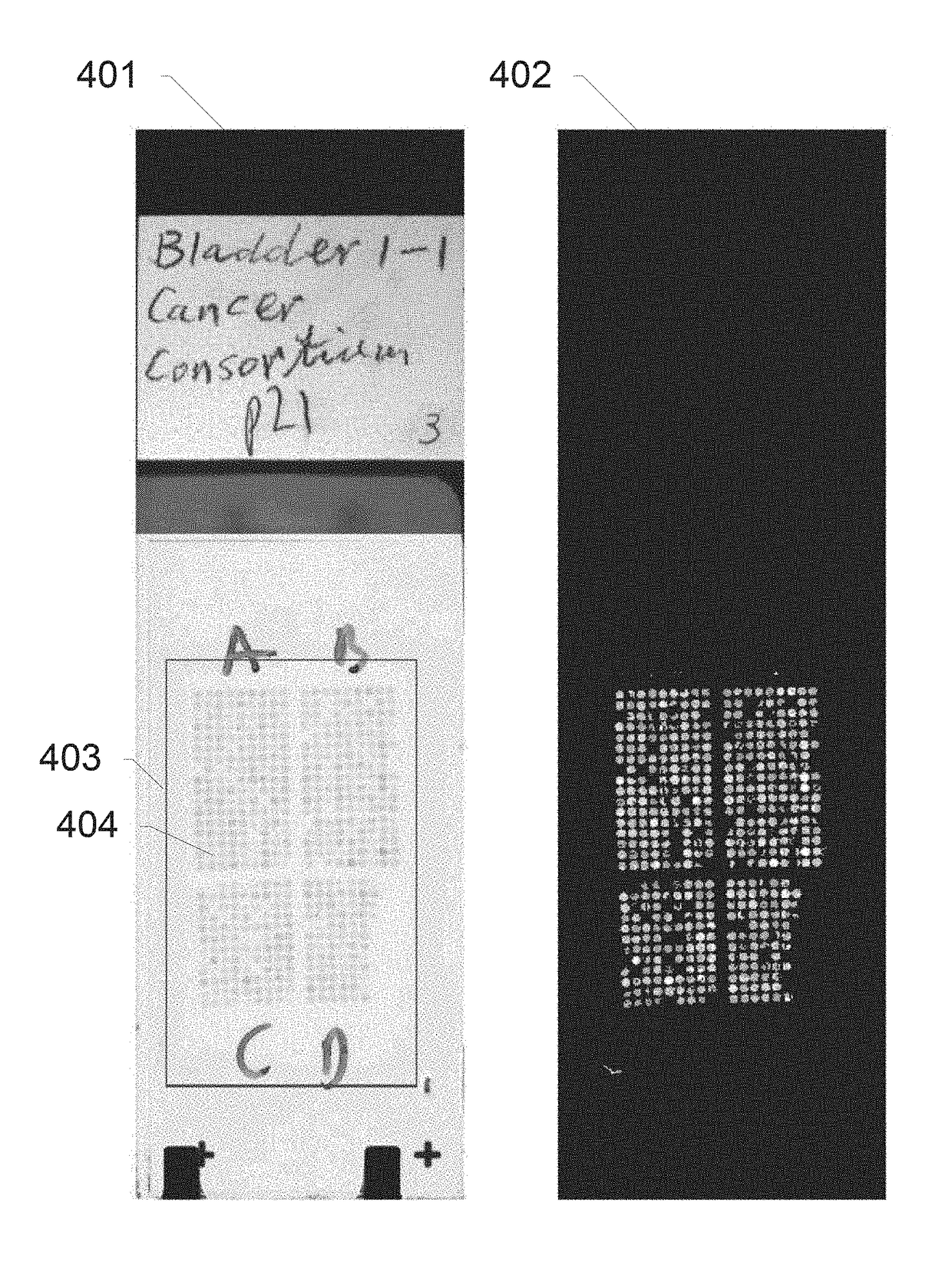
[0229]2. The image analysis system of embodiment 1, wherein the digital image is a thumbnail image.

[0230]3. The image analysis system of any one of the previous embodiments, wherein the image analysis system comprises a user interface, wherein the selection of the slide-type specific tissue detection routine comprises receiving a user's selection of a tissue slide type via the user interface; and selecting the one of the slide-type specific tissue detection routines assigned the tissue slide type selected by the user.

[0231]4. The image analysis system of any one of the previous embodiments, wherein the generic tissue detection routine and one or more of the slide-type specific tissue detection routines comprise a sub-routine for generating a binary mask (M) that selectively masks non-tissue regions, the sub-routine comprising: computing a histogram (800) of a grayscale version of the digital image; extracting a plurality of intensity thresholds (rts) from the histogram; applying the...

embodiment 4

[0232]5. The image analysis system of embodiment 4, the extraction of the plurality of intensity thresholds from the histogram comprising: identifying a max-grayscale-index (mgs), the max-grayscale-index being the maximum grayscale intensity value observed in the histogram; identifying a max-histogram-location (mhl), the max-histogram-location being the grayscale intensity value having the highest occurrence frequency in the histogram; computing a rightgap by subtracting the maximum-histogram-location (mhl)from the max-grayscale-index (mgs); computing a glass-left-cutoff according to glass-left-cutoff=max-histogram-location(mhl)-rightgap; performing the extraction of the plurality of thresholds such that the lowest one (dco[0]) of the thresholds is an intensity value equal to or larger than glass_left_cutoff−rightgap and such that the highest one of the thresholds is equal to or smaller than glass_left_cutoff+rightgap.

embodiment 5

[0233]6. The image analysis system of embodiment 5, the extraction of the plurality of intensity thresholds from the histogram comprising: computing an interval-range-gap(irg) according to irg=max(1, round(rightgap / constant)), wherein constant is a predefined numerical value between 2 and 20, preferentially between 3 and 8; computing a max_rightgap value according to max_rightgap=min(rightgap−1, round(rightgap * gap_cutoff_fraction), wherein the gap_cutoff_fraction is a predefined value in the range of 0.5-0.95, preferentially 0.7-0.8; computing a number_interval_terms according to number_interval_terms=((max_rightgap+rightgap) / interval_range_gap); creating the plurality of threshold such that their number is identical to the computed number_interval_terms.

[0234]7. The image analysis system of embodiments 4, 5 or 6, the application of the plurality of intensity thresholds (rts) on the digital image for generating a plurality of intermediate masks comprising: a) creating a first inte...


PUM


Abstract

The subject disclosure provides systems and methods for determination of Area of Interest (AOI) for different types of input slides. Slide thumbnails may be assigned into one of five different types, and separate algorithms for AOI detection executed depending on the slide type. Slide types include ThinPrep (RTM) slides, tissue micro-array (TMA) slides, control HER2 slides with 4 cores, smear slides, and a generic slide. The slide type may be assigned based on a user input. Customized AOI detection operations are provided for each slide type. If the user enters an incorrect slide type, operations include detecting the incorrect input and executing the appropriate method. The result of each AOI detection operations provides as its output a soft-weighted image having zero intensity values at pixels that are detected as not belonging to tissue, and higher intensity values assigned to pixels detected as likely belonging to tissue regions.

Description

CROSS-REFERENCE TO RELATED APPLICATIONS[0001]This patent application is a continuation of International Patent Application No. PCT / EP2016 / 051967 filed Jan. 29, 2016, which claims priority to and the benefit of U.S. Provisional Application No. 62 / 110,473, filed Jan. 31, 2015. Each of the above patent applications are hereby incorporated by reference herein in their entireties.BACKGROUND OF THE SUBJECT DISCLOSUREField of the Subject Disclosure[0002]The present subject disclosure relates to digital pathology. More particularly, the present subject disclosure relates to tissue detection on glass slides containing tissue biopsies used to generate whole slide scans.Background of the Subject Disclosure[0003]In the field of digital pathology, biological specimens such as tissue sections, blood, cell cultures and the like may be stained with one or more stains and analyzed by viewing or imaging the stained specimen. Observing the stained specimen, in combination with additional clinical info...

Claims


Application Information

Patent Timeline
Patent Type & Authority Applications(United States)
IPC IPC(8): G06T7/00G06F3/0482
CPCG06T7/0012G06T7/11G06T2207/10056G06T2207/30024G06F3/0482G06T2200/24G06T2207/20132G06T2207/30072G06T2207/20092G06V20/69
Inventor SARKAR, ANINDYAMARTIN, JIM
Owner VENTANA MEDICAL SYST INC
Why Eureka
Try Eureka