Self-Illuminated Handheld Lens for Retinal Examination and Photography and Related Method thereof

a handheld lens and self-illuminating technology, applied in the field of improvement, can solve the problems of significant optical aberrations that obscure retinal details, and achieve the effects of simple manufacturing, improved image quality, and improved image quality

Inactive Publication Date: 2018-03-01
UNIV OF VIRGINIA ALUMNI PATENTS FOUND
View PDF0 Cites 28 Cited by
  • Summary
  • Abstract
  • Description
  • Claims
  • Application Information

AI Technical Summary

Benefits of technology

[0016]An aspect of an embodiment of the present invention seeks to correct the illumination issues with current handheld fundus lenses. The slit lamp biomicroscope may be used to view the image through the handheld retina lens, but the handheld lens itself would provide the illumination of the retina, rather than relying on slit illumination provided by the slit lamp. An aspect of various embodiments of the present invention captures the illumination characteristics of retinal camera design in miniature, creating a design that may be battery powered and simple to manufacture, maintaining commercial viability. An embodiment provides, among other things, a handheld lens where ring illumination is directly coupled to the contact lens portion of the handheld lens, as illustrated in FIG. 3B. This creates a ring or “donut” of illumination that is transmitted to the eye when the front contact lens of the device is applied to the cornea. This ring could be created through many designs, including a ring of surface mount LEDs, the use of fiber optics to fashion donut illumination from a point source of light, the use of a shaped mirror to provide a dispersed donut of light, and a curved surface with surface mount LEDs providing multidirectional illumination. FIG. 3D illustrates the potential viewing area of such a widefield handheld contact imaging lens of FIG. 3B.
[0017]An aspect of an embodiment of the present invention provides a self-illuminated handheld lens (and related method) to aid in the examination and treatment of retinal disease. Retinal exams and retinal laser treatments are commonly performed using a slit lamp biomicroscope. The slit lamp uses a narrow beam of light that illuminates less than 1% of the total retinal surface and suffers from significant optical aberrations that obscure retinal details. To alleviate these problems, an embodiment of the present invention provides a self-illuminated handheld fundus lens for retinal imaging to be used in conjunction with the slit lamp biomicroscope to provide clear, reflection free, wide field illumination and view of the retina. An aspect of an embodiment of the present invention device creates ring illumination that is coupled through the contact lens of a handheld fundus lens to provide this widefield illumination of the retina. An embodiment of the present invention device may rely on use of fiber optics and / or light emitting diodes to provide illumination, though many other means of illumination are possible.

Problems solved by technology

The slit lamp uses a narrow beam of light that illuminates less than 1% of the total retinal surface and suffers from significant optical aberrations that obscure retinal details.

Method used


Image

  • Self-Illuminated Handheld Lens for Retinal Examination and Photography and Related Method thereof
  • Self-Illuminated Handheld Lens for Retinal Examination and Photography and Related Method thereof
  • Self-Illuminated Handheld Lens for Retinal Examination and Photography and Related Method thereof

Examples


Embodiment Construction

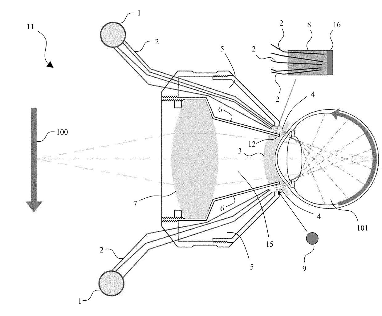
[0056]Aspects of the present invention are directed towards providing full and even illumination of a patient's retina through lighting integrated into a handheld fundus lens. By integrating the lighting, aspects of the present invention reduce and even eliminate many lens artifacts and reflections. By increasing the accuracy, quality, and field of view afforded during clinical examination of the retina, an aspect of the present invention will allow practitioners to make more accurate diagnoses and will increase safety during retinal surgical procedures.

[0057]An aspect of an embodiment of the current invention is designed to provide, among other things, self-contained ring illumination of the retina within the space constraints of a handheld fundus lens which measures approximately 40 mm×50 mm×20 mm, and optimized for visualization of retinal details by a trained practitioner at an ophthalmic slit lamp. As portability of the unit may be advantageous to marketability and acceptance o...


PUM


Abstract

System and method directed towards providing full and even illumination of a patient's retina through lighting integrated into a handheld fundus lens. By integrating the lighting, the method and system reduces and even eliminate many lens artifacts and reflections. By increasing the accuracy, quality, and field of view 10 afforded during clinical examination of the retina, the method and system will allow practitioners to make more accurate diagnoses and will increase safety during retinal surgical procedures.

Description

RELATED APPLICATIONS[0001]This application is a continuation-in-part (CIP) application of U.S. patent application Ser. No. 15 / 048,279, filed Feb. 19, 2016, which is a continuation-in-part (CIP) application of U.S. patent application Ser. No. 14 / 268,643, filed May 2, 2014, now U.S. Pat. No. 9,265,410, granted on Feb. 23, 2016, which is a division of U.S. patent application Ser. No. 13 / 318,695, filed Nov. 3, 2011, now U.S. Pat. No. 8,740,383, granted on Jun. 3, 2014, which is a national stage filing of International Application No. PCT / US2010 / 033875, filed May 6, 2010, which claims the benefit of priority under 35 U.S.C. §119(e) of U.S. Provisional Application Serial No. 61 / 175,807, filed on May 6, 2009, entitled “Self-Illuminated Handheld Lens for Retinal Examination and Photography and Related Method thereof;” the disclosures of which are hereby incorporated by reference herein in their entirety.FIELD OF THE INVENTION[0002]The present invention is directed to an improved device for ...

Claims


Application Information

Patent Timeline
Patent Type & Authority Applications(United States)
IPC IPC(8): A61B3/00H04N5/225G06K9/00A61B3/15A61B3/12A61B3/125A61B3/14
CPCH04N5/2256H04N5/2251G06K9/00604A61B3/156A61B3/1208A61B3/12A61B3/0008A61B3/125A61B3/14G06V40/19H04N23/56H04N23/50A61B3/135A61B3/0025
Inventor YATES, PAUL ANDREW
Owner UNIV OF VIRGINIA ALUMNI PATENTS FOUND
Why Eureka
Try Eureka