Auxiliary device for locating, mapping and microscopically measuring neoplasias

Inactive Publication Date: 2018-03-08
FUNDACAO ANTONIO PRUDENTE
View PDF1 Cites 2 Cited by
  • Summary
  • Abstract
  • Description
  • Claims
  • Application Information

AI Technical Summary

Benefits of technology

The patent describes a device used in medical procedures to hold and immobilize surgical specimens for study and analysis. The device consists of an upper and lower plate with a matrix of threads in between. The plates are pressed together to hold the specimen securely without damaging its physical structure. The device can be opened to allow greater contact between the specimen and an added formaldehyde solution, which improves the preservation of the specimen for further processing. The threads in the device leave a physical impression on the surface of the specimen, which helps to mark important areas of interest. This information is not lost when the specimen is removed from the device. Overall, the device provides a safe and effective way to hold and stabilize surgical specimens for analysis and study.

Problems solved by technology

The state of the art bears devices that are suited to just part of these needs, such as international application WO2014039727, but which are not suitable, for example, to contact with formaldehyde and to microscopic measurement.

Method used


Image

  • Auxiliary device for locating, mapping and microscopically measuring neoplasias
  • Auxiliary device for locating, mapping and microscopically measuring neoplasias
  • Auxiliary device for locating, mapping and microscopically measuring neoplasias

Examples


example

[0044]The following example, described with the aid of the accompanying drawings, is given as an illustration of a particular embodiment of the invention, without imposing restrictions thereon, other than those contained in the accompanying claims.

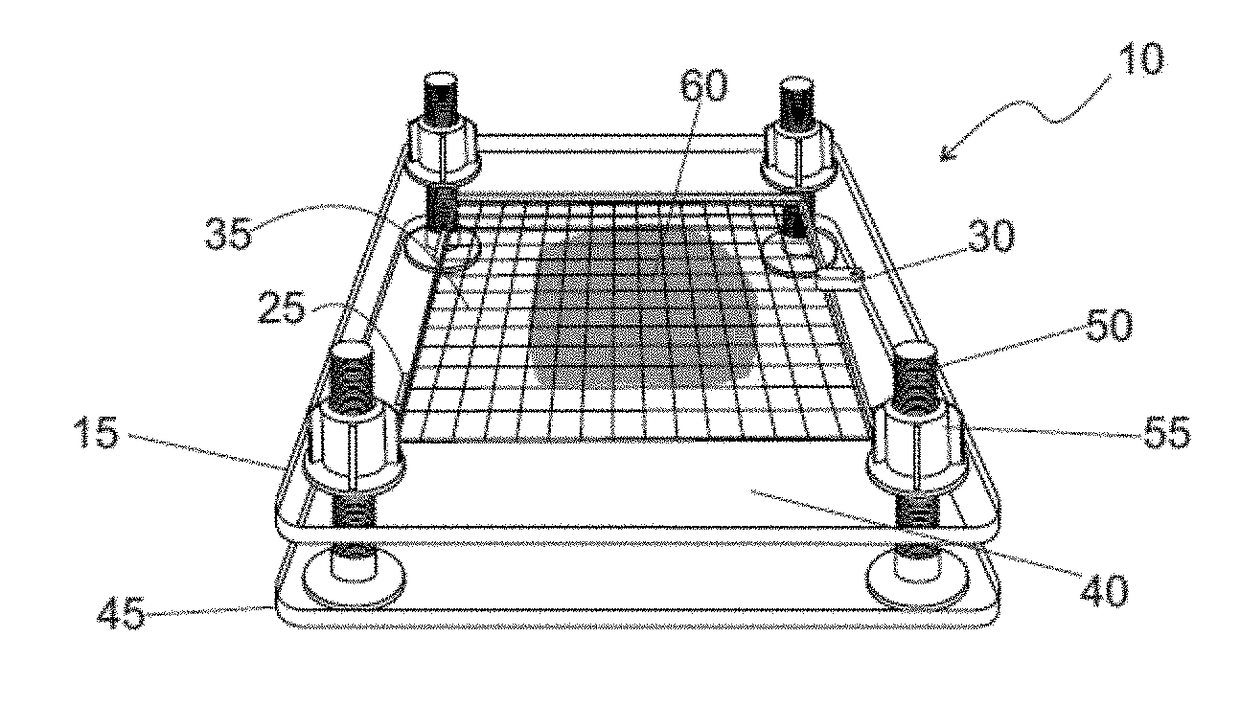
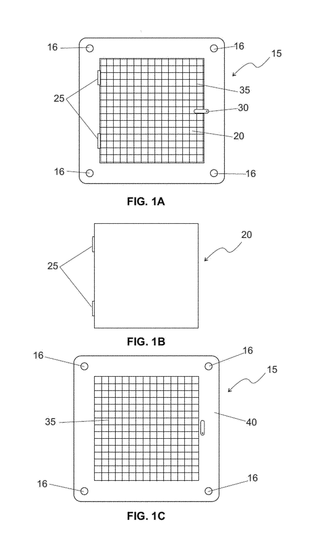
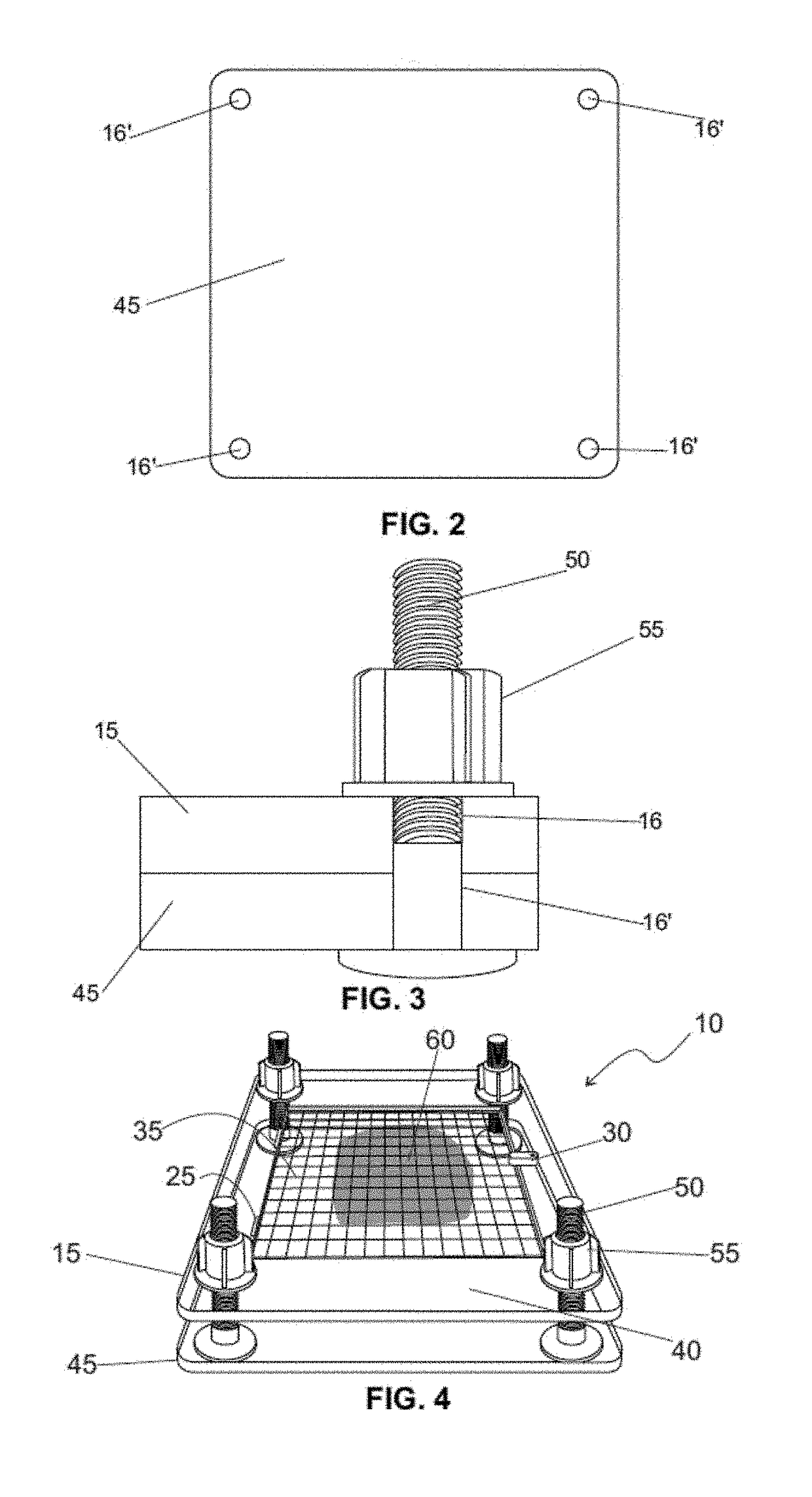
[0045]The device 10 of the invention comprises two cooperating plates, one upper plate 15 and one lower plate 45, with details presented in FIGS. 1A, 1B, 1C and 2. The dimensions of the plates are 21 cm×21 cm (width×length).

[0046]The upper plate 15 comprises two cooperating parts, namely a frame 40 and a tilting central cover 20 (measuring 15 cm×15 cm), said cover 20 cooperating with the frame 40 by way of hinges 25, and kept closed on a non-definitive basis by way of a lock 30.

[0047]The frame 40 and the central cover 20 of the upper plate 15 are made of radio-transparent acrylic, 10 mm and 5 mm in thickness, respectively.

[0048]A matrix 35 of fine nylon threads measuring 0.6 mm in diameter having low radio-opacity, is associated to the fra...


PUM


Abstract

The present invention refers to an auxiliary device for locating, mapping and microscopically measuring neoplasias, in particular microcalcifications in breast surgery specimens, linked to a radiological procedure.

Description

FIELD OF THE INVENTION[0001]The present invention refers to an auxiliary device for locating, mapping and microscopically measuring neoplasias, in particular microcalcifications in breast surgery specimens, linked to a radiological procedure.[0002]The device of the invention exerts a number of valuable functions, allowing a surgical specimen, the result of an excision:[0003](1) during the intraoperative period to be properly fixed to undergo radiography, allowing precise location of microcalcifications and decision on the need for surgical expansion,[0004](2) to be efficiently conserved by immersion in formaldehyde, in the same position and shape it was radiographed;[0005](3) to provide the mapping of the microcalcifications and microscopic measurement of tumor size for accurate pathological staging, depending on the radiographic quality and print of the matrix left in the specimen.BACKGROUND OF THE INVENTION[0006]Breast cancer accounts for 10% of all cancers diagnosed worldwide and...

Claims


Application Information

Patent Timeline
IPC IPC(8): G01N33/483A61B6/12G01N1/31G02B21/36G02B21/26
CPCG01N23/06G01N33/4833A61B6/12G01N1/312G02B21/365G02B21/26G01N23/08G01N1/286G01N1/36G01N23/083G01N23/20025
Inventor PEDAO, DIEGO MOREIRADE ANDRADE, VICTOR PIANA
Owner FUNDACAO ANTONIO PRUDENTE
Why Eureka
Try Eureka