Percutaneous delivery systems for anchoring an implant in a cardiac valve annulus

a technology of cardiac valve and implant, which is applied in the field of treatment of mitral or tricuspid valve prolapse and mitral regurgitation, can solve the problems of loss of ventricular function and geometry, insufficient durability of the valve, and inability to properly operate one or more of these valves, so as to facilitate proper leaflet coaptation

Active Publication Date: 2018-08-02
HEART REPAIR TECH INC
View PDF6 Cites 23 Cited by
  • Summary
  • Abstract
  • Description
  • Claims
  • Application Information

AI Technical Summary

Benefits of technology

[0027]In some embodiments, cinching comprises cinching the posterior annulus toward the anterior annulus. In some embodiments, cinching facilitates proper leaflet coaptation. In some embodiments, the first suture extends in a straight path through a pilot hole in the posterior annulus. In some embodiments, the second suture extends in a straight path through a pilot hole in the anterior annulus. In some embodiments, the method can include delivering a third subannular anchor coupled to a third suture, wherein the third suture extends through the annulus. In some embodiments, the method can include delivering a fourth subannular anchor coupled to a fourth suture, wherein the f...

Problems solved by technology

Various disease processes can impair the proper functioning of one or more of these valves.
In addition, damage to the ventricle from prior heart attacks (i.e., myocardial infarction secondary to coronary artery disease) or other heart diseases (e.g., cardiomyopathy) can distort the valve's geometry causing it to dysfunction.
The predominant cause of dysfunction of the mitral valve is regurgitation which produces an ineffective cardiac pump function resulting in several deleterious conditions such as ventricular and atrial enlargement, pulmonary hypertension and heart-failure and ultimately death.
Historically, the surgical approach to patients with functional mitral regurgitation was mitral valve replacement, ho...

Method used


Image

  • Percutaneous delivery systems for anchoring an implant in a cardiac valve annulus
  • Percutaneous delivery systems for anchoring an implant in a cardiac valve annulus
  • Percutaneous delivery systems for anchoring an implant in a cardiac valve annulus

Examples


Embodiment Construction

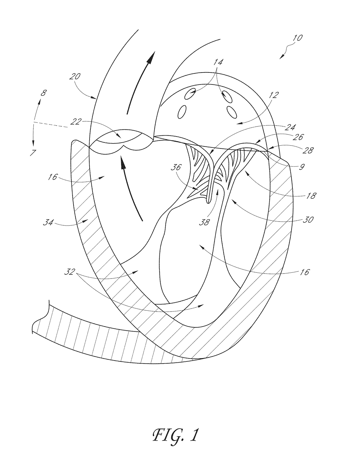
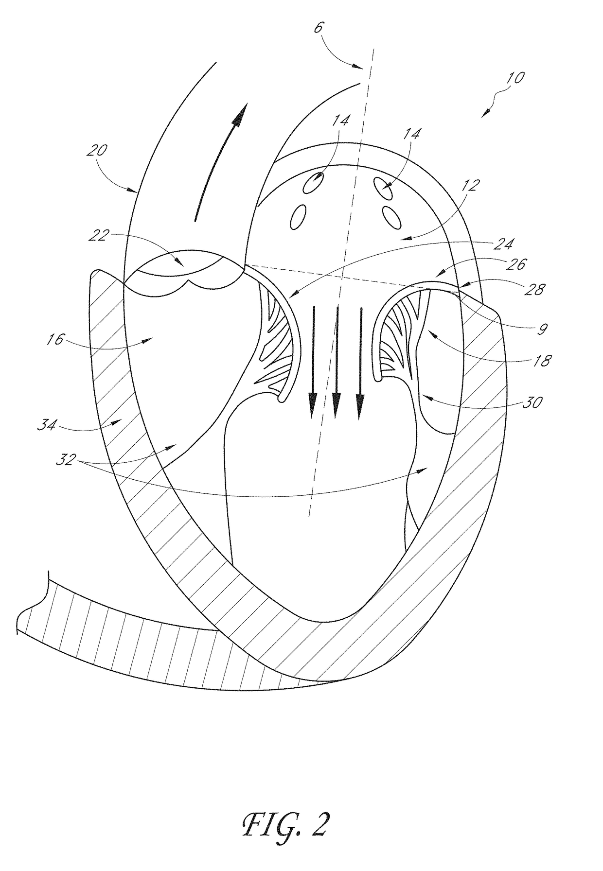
[0144]FIG. 1 illustrates a cross-sectional view of the heart 10 with a normal mitral valve 18 in systole. As illustrated, the heart 10 comprises the left atrium 12 which receives oxygenated blood from the pulmonary veins 14 and the left ventricle 16 which receives blood from the left atrium 12. The mitral valve 18 is located between the left atrium 12 and left ventricle 16 and functions to regulate the flow of blood from the left atrium 12 to the left ventricle 16. During ventricular diastole, the mitral valve 18 is open which allows blood to fill the left ventricle 16. During ventricular systole, the left ventricle 16 contracts, which results in an increase in pressure inside the left ventricle 16. The mitral valve 18 closes when the pressure inside the left ventricle 16 increases above the pressure within the left atrium 12. The pressure within the left ventricle 16 continues increasing until the pressure within the left ventricle 16 exceeds the pressure within the aorta 20, which...


PUM


Abstract

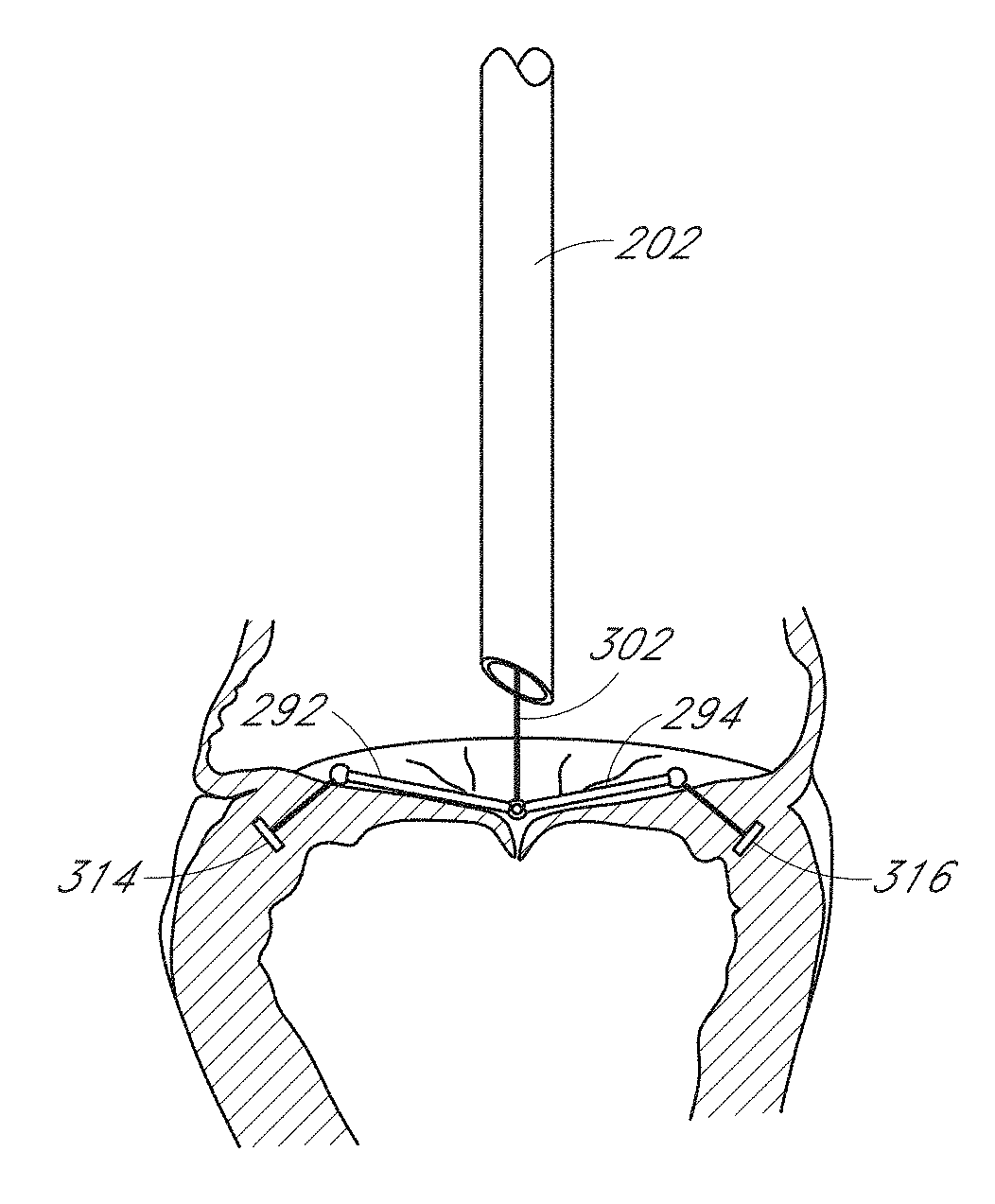
Mitral valve prolapse and mitral regurgitation can be treating by implanting in the mitral annulus a transvalvular intraannular band. The band has a first end, a first anchoring portion located proximate the first end, a second end, a second anchoring portion located proximate the second end, and a central portion. The central portion is positioned so that it extends transversely across a coaptive edge formed by the closure of the mitral valve leaflets. The band may be implanted via translumenal access or via thoracotomy.

Description

CROSS-REFERENCE TO RELATED APPLICATIONS[0001]This application claims the benefit under 35 U.S.C. § 119(e) as a nonprovisional application of U.S. Prov. App. No. 62 / 437,898 filed on Dec. 22, 2016, U.S. Prov. App. No. 62 / 491,750 filed Apr. 28, 2017, and U.S. Prov. App. No. 62 / 549,215 filed Aug. 23, 2017. The disclosure of each of the aforementioned priority applications is hereby incorporated by reference herein in their entireties. This application is also related to U.S. application Ser. No. 15 / 293111, filed Oct. 13, 2016, which in turn claims the benefit under 35 U.S.C. § 119(e) as a nonprovisional application of U.S. Prov. App. No. 62 / 241,687 filed on Oct. 14, 2015. This application is also related to U.S. patent application Ser. No. 14 / 628,114 filed on Feb. 20, 2015, which is in turn a continuation of U.S. patent application Ser. No. 13 / 650,998 filed Oct. 12, 2012, now issued as U.S. Pat. No. 8,961,597 on Feb. 24, 2015, which is a continuation of U.S. patent application Ser. No. ...

Claims


Application Information

Patent Timeline
IPC IPC(8): A61F2/24A61B18/14
CPCA61M2025/0095A61M25/0026A61B2018/00369A61B2018/00577A61B2018/00589A61B17/0469A61F2/2457A61F2/2466A61F2230/005A61F2/246A61F2/2445A61B18/1492A61F2230/0006A61F2230/0039A61F2230/0036A61F2230/001A61F2230/0013A61F2230/0008A61F2230/0023A61B2017/00247A61B2017/003A61F2220/0008A61B2018/00607A61B17/0401A61B17/0482A61B2017/00243A61B2017/00783A61B2017/00986A61B2017/0406A61B2017/0409A61B2017/0417A61B2017/0419A61B2017/0464A61B2017/06052A61B2017/0647A61B2017/0649A61F2/2454
Inventor SUBRAMANIAN, VALAVANUR A.REO, MICHAEL L.HULME, GARYAFZAL, THOMASCHRISTIAN, JEFFREYBUCHBINDER, MAURICE
Owner HEART REPAIR TECH INC
Why Eureka
Try Eureka