Method and System for Dose-Optimized Computed Tomography Scanning of a Target Organ

a computed tomography and target organ technology, applied in the field of planning and acquisition of dose-optimized ct lung scans, to achieve the effect of dose-optimized ct lung scan acquisition

Active Publication Date: 2018-08-16
SIEMENS HEALTHCARE GMBH
View PDF0 Cites 17 Cited by
  • Summary
  • Abstract
  • Description
  • Claims
  • Application Information

AI Technical Summary

Benefits of technology

[0004]The present invention provides a method and system for dose-optimized CT lung scan acquisition. Embodiments of the present invention provide a fully automated dose-optimized CT lung scan acquisition without the need for a tomogram acquisition and without in...

Problems solved by technology

CT scans expose patients to...

Method used


Image

  • Method and System for Dose-Optimized Computed Tomography Scanning of a Target Organ
  • Method and System for Dose-Optimized Computed Tomography Scanning of a Target Organ
  • Method and System for Dose-Optimized Computed Tomography Scanning of a Target Organ

Examples


Embodiment Construction

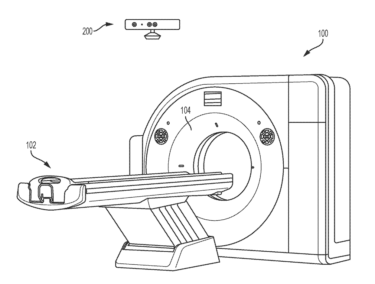
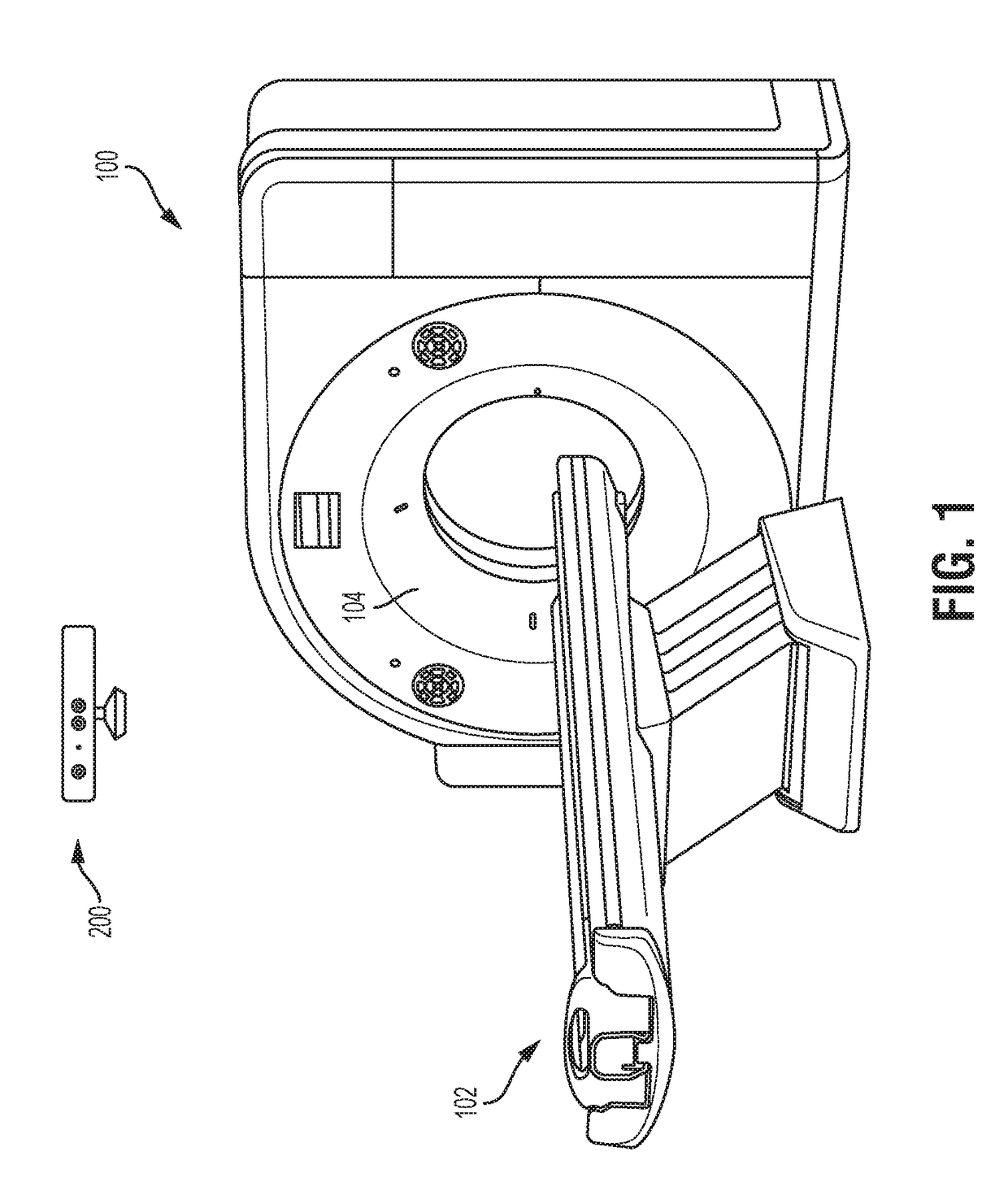
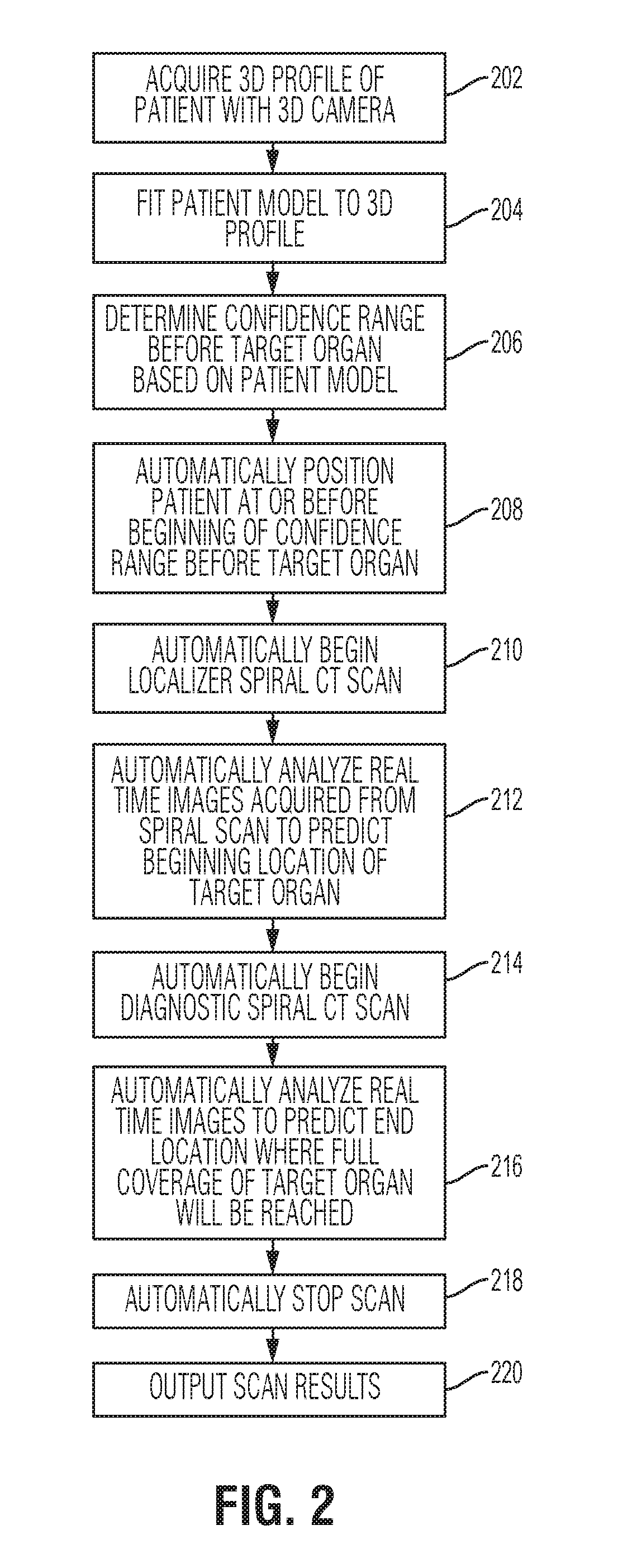
[0011]The present invention relates to a method and system for dose-optimized acquisition of a CT scan of a target organ. Embodiments of the present invention are described herein to give a visual understanding of the methods for scan range determination and CT lung scan acquisition. A digital image is often composed of digital representations of one or more objects (or shapes). The digital representation of an object is often described herein in terms of identifying and manipulating the objects. Such manipulations are virtual manipulations accomplished in the memory or other circuitry / hardware of a computer system. Accordingly, is to be understood that embodiments of the present invention may be performed within a computer system using data stored within the computer system.

[0012]Lung scans currently require topographic scans of the patient previous to the actual acquisition of the diagnostic CT lung scan. Such a topographic scan is used to obtain a topogram, which is a scout proje...


PUM


Abstract

A method and system for dose-optimized acquisition of a computed tomography (CT) scan of a target organ is disclosed. A localizer spiral CT scan is started at a beginning of a confidence range before a target organ. Real-time localizer scan images are automatically analyzed to predict a beginning location of the target organ based on the real-time localizer scan images. A diagnostic spiral CT scan is automatically started at the predicted beginning location of the target organ. Real-time diagnostic scan images are automatically analyzed to predict an end location of the target organ where full coverage of the target organ will be reached. The diagnostic spiral CT scan is automatically stopped in response to reaching the predicted end location of the target organ. A 3D profile can be acquired using a 3D camera and used to determine the confidence range before the target organ.

Description

BACKGROUND OF THE INVENTION[0001]The present invention relates to planning and acquisition of computed tomography (CT) scans of a target organ, and more particularly, to planning and acquisition of dose-optimized CT lung scans.[0002]Computed tomography (CT) is a medical imaging technique that makes use of computer-processed combinations of many X-ray projection images taken from different angles to produce cross-sectional images of specific areas of a scanned object. CT can be used to generate a three-dimensional (3D) image of the inside of the scanned object from a series of two-dimensional projection images taken around a single axis of rotation. CT chest or lung scans can be used for detecting both acute and chronic changes in the lung parenchyma. For example, because CT is able to detect very small nodules in the lung, CT lung scans are often used for diagnosing lung cancer at its earliest, most curable stage.[0003]CT scans expose patients to radiation, which can be harmful. Lun...

Claims


Application Information

Patent Timeline
IPC IPC(8): A61B6/03A61B6/04A61B6/00
CPCA61B6/032A61B6/5205A61B6/50A61B6/0457A61B6/027A61B6/488A61B6/5217A61B6/542A61B6/545G16H50/30A61B6/0487
Inventor VEGA, FERNANDOSAUER, FRANK
Owner SIEMENS HEALTHCARE GMBH
Why Eureka
Try Eureka