Deep Convolutional Encoder-Decoder for Prostate Cancer Detection and Classification

Active Publication Date: 2018-08-23
SIEMENS HEALTHCARE GMBH
View PDF0 Cites 32 Cited by
  • Summary
  • Abstract
  • Description
  • Claims
  • Application Information

AI Technical Summary

Benefits of technology

The present invention provides a method and system for automatically detecting and classifying prostate tumors in MRIs. This is done by using a deep learning network that can directly detect and classify tumors in the MRI images, without needing to extract features from the images first. The system receives a set of multi-parametric MRI images, including different types of MRI images, and detects and classifies prostate tumors in all of them simultaneously. This approach helps to improve the accuracy and efficiency of identifying prostate tumors in MRI images.

Problems solved by technology

Manual reading multi-parametric MR images, which consist of as many as eight image channels, can be a tedious task.
Furthermore, subtle and collective signatures of a cancerous lesion expressed within multi-parametric MR images are difficult to detect, even by an expert radiologist.

Method used


Image

  • Deep Convolutional Encoder-Decoder for Prostate Cancer Detection and Classification
  • Deep Convolutional Encoder-Decoder for Prostate Cancer Detection and Classification
  • Deep Convolutional Encoder-Decoder for Prostate Cancer Detection and Classification

Examples


Embodiment Construction

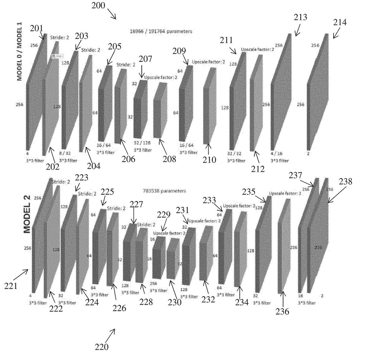
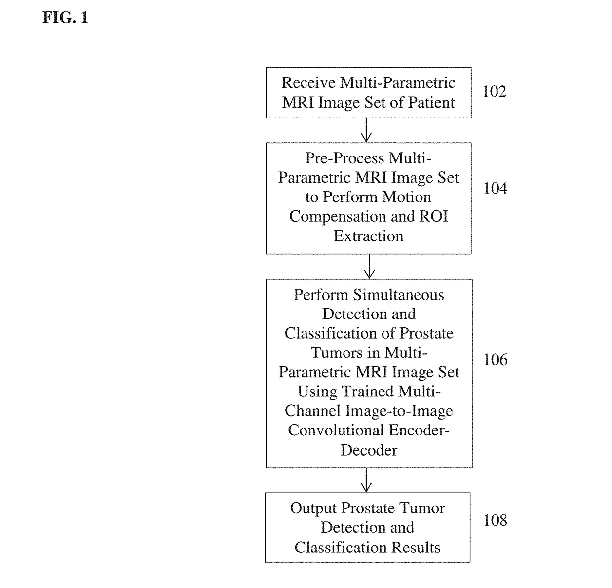
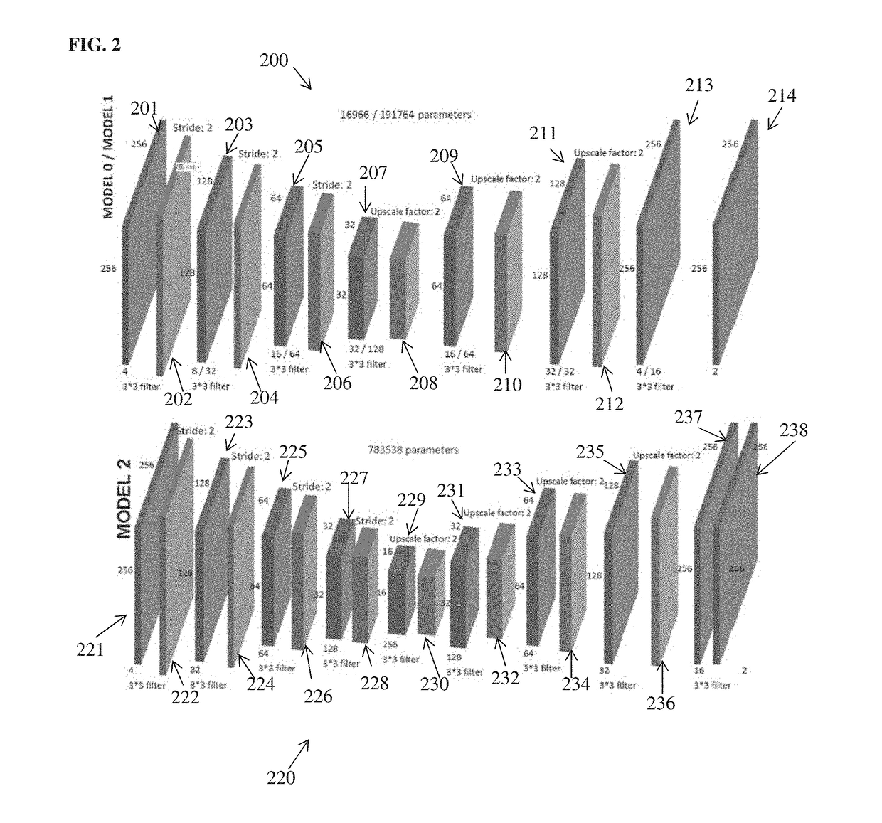
[0016]The present invention relates to a method and system for automated computer-based detection and classification of prostate tumors in multi-parametric magnetic resonance (MR) images. Embodiments of the present invention are described herein to give a visual understanding of the method for automated detection and classification of prostate tumors. A digital image is often composed of digital representations of one or more objects (or shapes). The digital representation of an object is often described herein in terms of identifying and manipulating the objects. Such manipulations are virtual manipulations accomplished in the memory or other circuitry / hardware of a computer system. Accordingly, is to be understood that embodiments of the present invention may be performed within a computer system using data stored within the computer system.

[0017]Various techniques have been proposed to provide an automated solution to detection and classification of prostate cancer using multi-...


PUM


Abstract

A method and apparatus for automated prostate tumor detection and classification in multi-parametric magnetic resonance imaging (MRI) is disclosed. A multi-parametric MRI image set of a patient, including a plurality of different types of MRI images, is received. Simultaneous detection and classification of prostate tumors in the multi-parametric MRI image set of the patient are performed using a trained multi-channel image-to-image convolutional encoder-decoder that inputs multiple MRI images of the multi-parametric MRI image set of the patient and includes a plurality of output channels corresponding to a plurality of different tumor classes. For each output channel, the trained image-to image convolutional encoder-decoder generates a respective response map that provides detected locations of prostate tumors of the corresponding tumor class in the multi-parametric MRI image set of the patient.

Description

[0001]This application claims the benefit of U.S. Provisional Application No. 62 / 461,970, filed Feb. 22, 2017, the disclosure of which is herein incorporated by reference.BACKGROUND OF THE INVENTION[0002]The present invention relates to automated detection and classification of prostate tumors in medical images, and more particularly, to automated detection and classification of prostate tumors in multi-parametric magnetic resonance (MR) images using a deep learning network.[0003]Prostate cancer is the most frequently diagnosed cancer in American men with 181,000 new cases in 2016 resulting in more than 26,000 deaths. Early diagnosis often results in long term survival, but depends on invasive multicore biopsies that are typically guided by transrectal ultrasound (TRUS) imaging. Recently, multi-parametric magnetic resonance imaging (MRI) has shown promising results for use in non-invasive prostate cancer detection, as a strong correlation has been established between mpMRI and histo...

Claims


Application Information

Patent Timeline
IPC IPC(8): G06T7/00
CPCG06T7/0012G06T2207/10096G06T2207/20081G06T2207/30096G06T2207/20084G06T9/001G06T9/002G06T2207/10088G06T2207/30081
Inventor KIRALY, ATILLA PETERABI NADER, CLEMENT JADGRIMM, ROBERTKIEFER, BERTHOLDKAMEN, ALI
Owner SIEMENS HEALTHCARE GMBH
Why Eureka
Try Eureka