Systems, methods, and apparatuses for digital histopathological imaging for prescreened detection of cancer and other abnormalities

a digital histopathological and imaging technology, applied in the field of digital histopathological imaging, can solve the problems of wasting time, wasting resources, and wasting resources, and achieve the effect of reducing the or apparatus, and solving the long-felt but unresolved need for a system, method or apparatus

Inactive Publication Date: 2018-09-06
INSPIRATA INC
View PDF6 Cites 17 Cited by
  • Summary
  • Abstract
  • Description
  • Claims
  • Application Information

AI Technical Summary

Benefits of technology

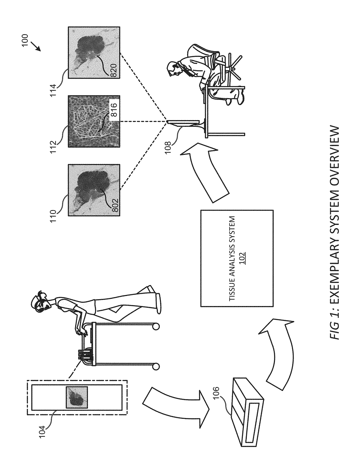
[0006]Histopathology images generally comprise digitized versions of microscopic views of tissue slides that may contain pieces of tissue from various human organs or abnormal masses within the body (e.g., lymph node, tumor, skin, etc.). These histopathology images may be collected for review by a trained professional (e.g., pathologist) to diagnose a particular disease, determine whether a tumor is malignant or benign, determine whether a surgeon has completely excised a tumor, etc. To expedite the review process (because pathologists view hundreds of histopathology images daily and often make only a binary decision regarding a particular histopathology image: yes, the image contains cancerous cells or, no, the image does not contain cancerous cells), the tissue analysis system described in the present disclosure processes histopathology images to identify / highlight regions of interest (e.g., a region that may comprise parts of a tumor, cancerous cells, or other predetermined abnormality), typically for subsequent review by a pathologist or other trained professional. Generally, by identifying / highlighting regions of interest within the histopathology images, the pathologist's review time of a particular histopathology image is reduced because the review need only cover the regions of interest and not the entire histopathology image. In some embodiments, professionals need not review the results of the processing because the tissue analysis system automatically identifies abnormalities within the histopathology images.
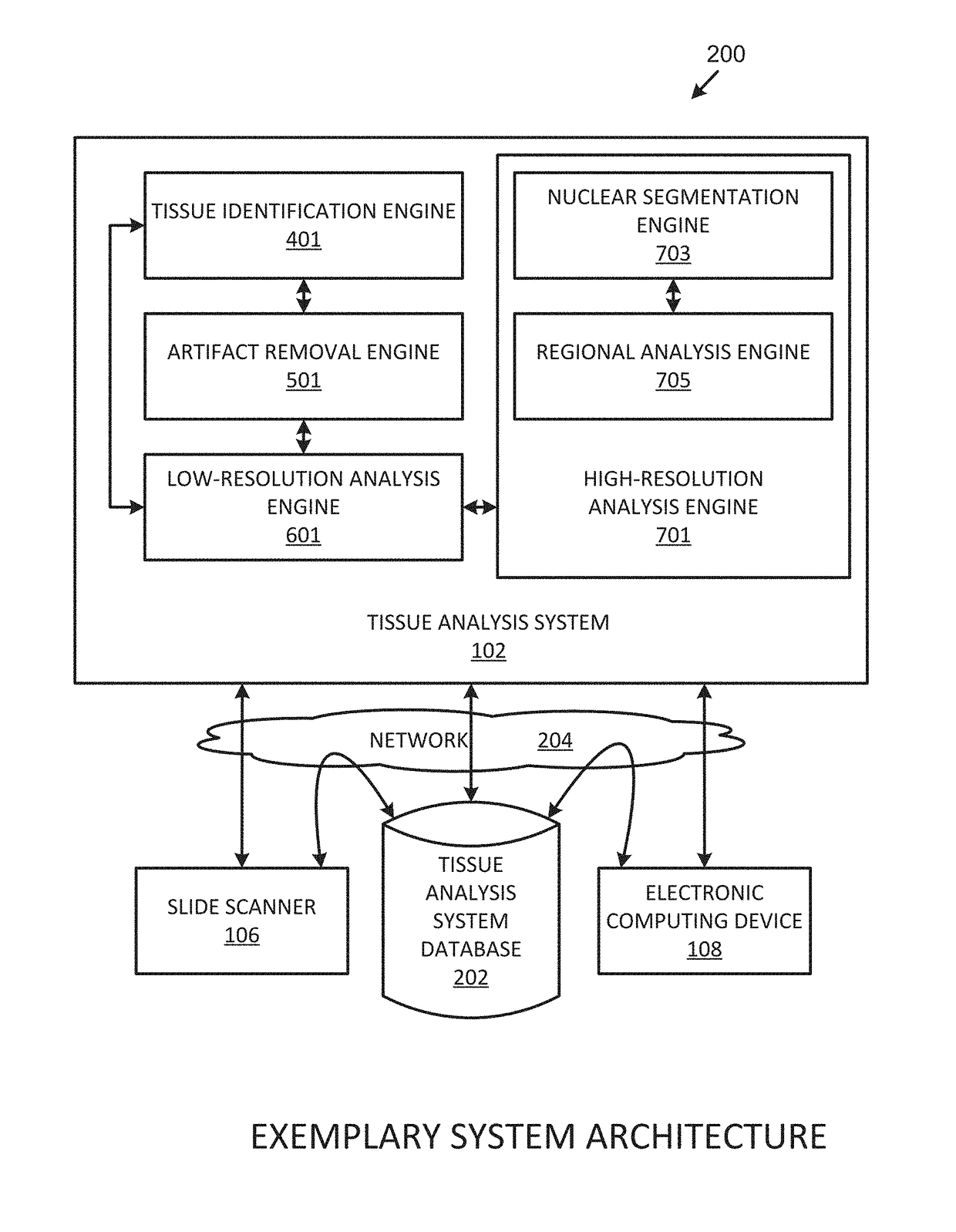
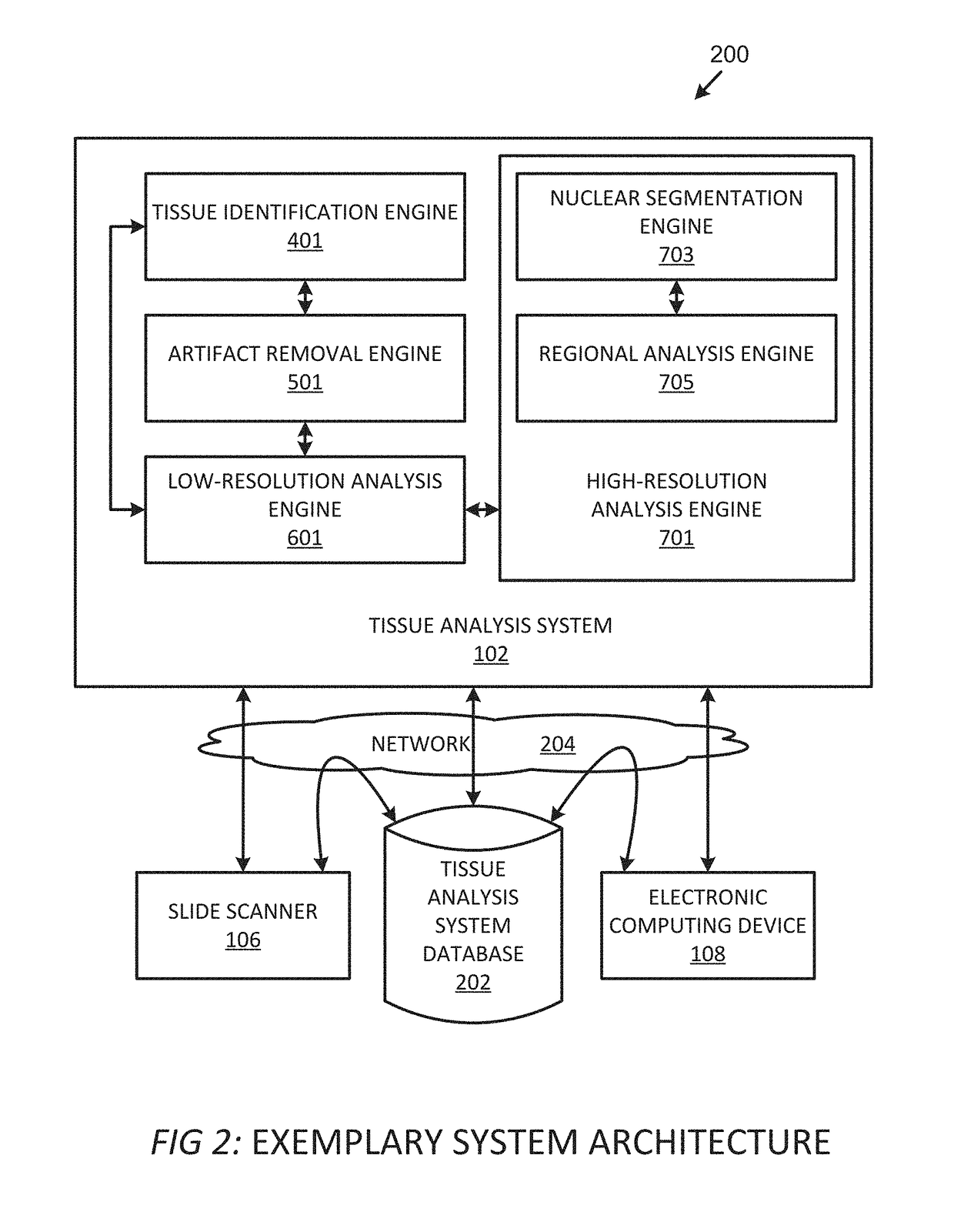
[0008]For example, to determine whether a patient has cancer, the tissue analysis system may process the histopathology image(s) of lymph node tissue to identify regions of interest that may contain cancerous cells. Accordingly, the histopathology image(s) undergo the tissue identification process to identify the lymph node tissue within the histopathology image(s) and confirm that the tissue is lymph node tissue and not some other tissue (e.g., adipose tissue, etc.). Similarly, the histopathology image(s) undergo the artifact removal process to remove any artifacts contained within the histopathology image(s). The histopathology image(s) undergo the low-resolution analysis process to quickly identify potential regions of interest for further analysis during the high-resolution analysis process, and the high-resolution analysis process, during which the tissue analysis system identifies / flags, for subsequent review by a pathologist, regions of interest that may contain cancerous cells. Thus, the pathologist may quickly review the histopathology image(s) of the lymph node tissue to determine whether a patient has cancer.

Problems solved by technology

Anytime a pathologist reviews a histopathology image, the pathologist must review the entire image to determine whether a particular abnormality is present, which can be a slow and tedious process.
In some cases, a resident physician is assigned the task of initially reviewing histopathology images to identify potential abnormalities for review by the pathologist, which also can further delay the histopathology review process.
Therefore, there is a long-felt but unresolved need for a system, method, or apparatus that quickly and efficiently analyzes histopathology images to determine the presence of certain predetermined abnormalities.

Method used


Image

  • Systems, methods, and apparatuses for digital histopathological imaging for prescreened detection of cancer and other abnormalities
  • Systems, methods, and apparatuses for digital histopathological imaging for prescreened detection of cancer and other abnormalities
  • Systems, methods, and apparatuses for digital histopathological imaging for prescreened detection of cancer and other abnormalities

Examples


Embodiment Construction

[0033]For the purpose of promoting an understanding of the principles of the present disclosure, reference will now be made to the embodiments illustrated in the drawings and specific language will be used to describe the same. It will, nevertheless, be understood that no limitation of the scope of the disclosure is thereby intended; any alterations and further modifications of the described or illustrated embodiments, and any further applications of the principles of the disclosure as illustrated therein are contemplated as would normally occur to one skilled in the art to which the disclosure relates. All limitations of scope should be determined in accordance with and as expressed in the claims.

[0034]Whether a term is capitalized is not considered definitive or limiting of the meaning of a term. As used in this document, a capitalized term shall have the same meaning as an uncapitalized term, unless the context of the usage specifically indicates that a more restrictive meaning f...


PUM

PropertyMeasurementUnit
diameteraaaaaaaaaa
lengthaaaaaaaaaa
brightnessaaaaaaaaaa

Abstract

Systems, methods, and apparatuses for analyzing histopathology images to determine the presence of certain predetermined abnormalities. The system processes histopathology images to identify / highlight regions of interest (e.g., a region that may comprise parts of a tumor, cancerous cells, or other predetermined abnormality) for subsequent review by a pathologist or other trained professional. For example, the system may process histopathology images of H&E-stained lymph node tissue to identify potentially cancerous cells within the histopathology images.

Description

CROSS REFERENCE TO RELATED APPLICATIONS[0001]This application claims priority to, the benefit under 35 U.S.C. § 119 of, and incorporates by reference herein in its entirety U.S. Provisional Patent Application No. 62 / 136,051, filed Mar. 20, 2015, and entitled “Systems, Methods, and Apparatuses for Digital Whole Slide Imaging for Prescreened Detection of Cancer and Other Abnormalities.”TECHNICAL FIELD[0002]The present systems, methods, and apparatuses relate generally to digital histopathological imaging and, more particularly, to analyzing histopathology images to determine the presence of certain predetermined abnormalities.BACKGROUND[0003]Pathologists review histopathology images to determine whether any abnormalities are present in the tissue within the image. For example, a pathologist may review histopathology images of tissue collected during a visit to a dermatologist to determine whether a mole is a carcinoma. Or, a pathologist may review tissue collected from a patient durin...

Claims


Application Information

Patent Timeline
Patent Type & Authority Applications(United States)
IPC IPC(8): G06K9/00G01N33/483G06K9/46G06T7/00G06V10/56
CPCG06K9/00147G01N33/4833G06K9/4652G06T7/0012G06T2207/30024G01F19/00G06V20/698G06V10/56G06V2201/031
Inventor LLOYD, MARK C.MONACO, JAMESVERMA, NISHANTHARDING, DAVID SCOTTMONTEAGUDO, MAYKEL OROZCOGOSSAGE, KIRK WILLIAMBABU, JANANI SIVASANKAR
Owner INSPIRATA INC
Why Eureka
Try Eureka