Digital pathology system

a digital pathology and pathology technology, applied in the field of digital pathology, can solve the problems of difficult automatic registration by computer algorithms, the readability of digital data, etc., and achieve the effect of improving the workflow

Active Publication Date: 2018-09-20
KONINKLJIJKE PHILIPS NV
View PDF3 Cites 10 Cited by
  • Summary
  • Abstract
  • Description
  • Claims
  • Application Information

AI Technical Summary

Benefits of technology

[0003]There may be a need to provide a system for improving the workflow in the process of selecting a region of interest of an unstained sample to be removed for molecular diagnostic.

Problems solved by technology

The weak contrast may affect the readability of the digital images and thus make it difficult for e.g. automatic registration by a computer algorithm.

Method used


Image

  • Digital pathology system
  • Digital pathology system
  • Digital pathology system

Examples


Embodiment Construction

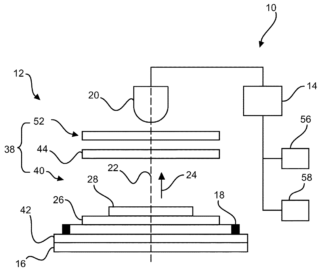
[0070]FIG. 1 shows an example of a system 10 for use in digital pathology. The system 10 comprises an image forming device 12 and a registration device 14. The image forming device 12 comprises a light source 16, an object receiving arrangement 18 (not shown in detail) and an image detector 20. The light source and the image detector are arranged in an optical path 22 (indicated with a dashed line). The dashed line is for illustration purpose only and forms no part of the claimed invention.

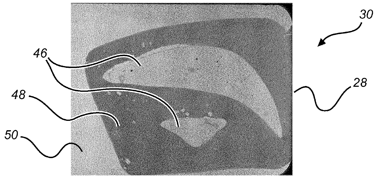
[0071]The light source 16 is configured to provide light 24 (indicated with an arrow) passing through a sample slide 26 to be received by the image detector 20. The object receiving arrangement 18 is configured to receive a sample slide 26 with a sample slice 28 of an object comprising unstained biological material and to position the sample slice 28 in the optical path 22 for acquiring a digital sample image 30 (not shown in FIG. 1, see examples in FIGS. 2B and 3B) of the sample slice 28. The reg...


PUM

PropertyMeasurementUnit
thicknessaaaaaaaaaa
thicknessaaaaaaaaaa
angleaaaaaaaaaa

Abstract

The present invention relates to digital pathology. In order to improve the workflow in the process of selecting a region of interest of an unstained sample to be removed for molecular diagnostic, a method (100) is provided for selecting a sample removing area of an unstained sample to be removed for molecular diagnostic. The method comprises the following steps: In a first step 102, also referred to as step a), a reference removing area is selected in a reference image of a reference slice of an object, wherein biological material in the reference slice is stained. In a second step 104, also referred to as step b), a digital sample image of a sample slice of the object is obtained under an imaging setting. The biological material in the sample slice is unstained. The sample slice is received on a sample slide and positioned in an optical path between a light source and an image detector. In the optical path between the light source and the image detector, it is further provided a contrast enhancing arrangement for improving contrast between the unstained biological material and background. Light is provided passing through the sample slice to be received by the image detector. In a third step 106, also referred to as step c), the digital sample image is registered with the reference image for translating the reference removing area in the reference image to the digital sample image. In a fourth step 108, also referred to as step d), a sample removing area is identified in the digital sample image based on the translated reference removing area.

Description

FIELD OF THE INVENTION[0001]The present invention relates to the field of digital pathology, and in particular to a system used in digital pathology, to a method for integrating digital pathology with molecular diagnostics. Furthermore, the invention relates to a computer program element as well as to a computer-readable medium.BACKGROUND OF THE INVENTION[0002]Digital Pathology refers to the creation, viewing, management, sharing, analysis, and interpretation of digital images of sample slides comprising biological material (e.g. tissues) and includes workflow considerations unique to a digital imaging environment. An important performance aspect of digital pathology is the quality of the digital images. For example, some digital images may have a weak contrast, when the sample slides comprise unstained biological material, such as unstained tissue. The weak contrast may affect the readability of the digital images and thus make it difficult for e.g. automatic registration by a comp...

Claims


Application Information

Patent Timeline
Patent Type & Authority Applications(United States)
IPC IPC(8): G02B21/36G01N1/30G02B21/00G02B21/12G02B21/34
CPCG02B21/367G01N1/30G02B21/0068G02B21/12G02B21/34G06T3/00G02B21/36
Inventor BOAMFA, MARIUS IOSIFPIERIK, ANKEWIMBERGER-FRIEDL, REINHOLDVAN LEEUWEN, MARINUS BASTIAAN (RIEN)
Owner KONINKLJIJKE PHILIPS NV
Why Eureka
Try Eureka