Device and method for spatiotemporal reconstruction of a moving vascular pulse wave in the brain and other organs

a vascular pulse wave and spatiotemporal reconstruction technology, applied in image enhancement, instruments, angiography, etc., can solve the problem that the equivalent volume of blood that moves to the brain with each heart beat has a measurable mortality

Active Publication Date: 2019-02-14
ANGIOWAVE IMAGING LLC
View PDF2 Cites 11 Cited by
  • Summary
  • Abstract
  • Description
  • Claims
  • Application Information

AI Technical Summary

Benefits of technology

The patent describes a technique for extracting cardiac frequency phenomena from an angiographic study of the brain. The technique involves acquiring raw data in the form of a brain angiogram at a much higher than cardiac frequency. This is done using ultrasound angiography, optical angiography, or fluoroscopic angiography. The technique involves performing a wavelet transform on the angiographic data, filtering for cardiac frequency, and then performing an inverse wavelet transform to render each frame as an image with a brightness hue color model. The technique can be used to study the vascular pulse wave in the brain and can provide valuable information for diagnostic and therapeutic purposes.

Problems solved by technology

Yet the equivalent volume of blood that moves to the brain with each heart beat has a measurable mortality if freely released into the cranial cavity.

Method used


Image

  • Device and method for spatiotemporal reconstruction of a moving vascular pulse wave in the brain and other organs
  • Device and method for spatiotemporal reconstruction of a moving vascular pulse wave in the brain and other organs
  • Device and method for spatiotemporal reconstruction of a moving vascular pulse wave in the brain and other organs

Examples


Embodiment Construction

[0025]The disclosure will now be described with reference to the drawing figures, in which like reference numerals refer to like parts throughout. Aspects of the disclosure advantageously provide a method and device for spatiotemporal reconstruction of a moving vascular pulse wave in the brain.

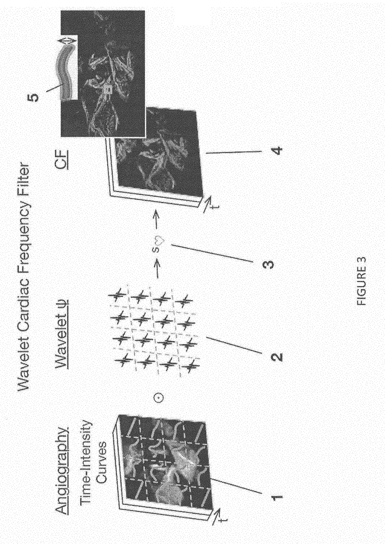
[0026]The brain appears to have organized cardiac frequency angiographic phenomena with such coherence as to qualify as vascular pulse waves. Separate arterial and venous vascular pulse waves may be resolved. This disclosure states the method of extracting a spatiotemporal reconstruction of the cardiac frequency phenomena present in an angiogram obtained at faster than cardiac frequency. A wavelet transform is applied to each of the pixel-wise time signals of the angiogram. The cardiac frequency phenomena are extracted by setting to zero all wavelet coefficients except for those corresponding to cardiac wavelet scale. The result is inverse wavelet transformed pixel-wise to give a spatiotempora...


PUM


Abstract

The brain appears to have organized cardiac frequency angiographic phenomena with such coherence as to qualify as vascular pulse waves. Separate arterial and venous vascular pulse waves may be resolved. This disclosure states the method of extracting a spatiotemporal reconstruction of the cardiac frequency phenomena present in an angiogram obtained at faster than cardiac frequency. A wavelet transform is applied to each of the pixel-wise time signals of the angiogram. If there is motion alias then instead a high frequency resolution wavelet transform of the overall angiographic time intensity curve is cross-correlated to high temporal resolution wavelet transforms of the pixel-wise time signals. The result is filtered for cardiac wavelet scale then pixel-wise inverse wavelet transformed. This gives a complex-valued spatiotemporal grid of cardiac frequency angiographic phenomena. It may be rendered with a brightness-hue color model or subjected to further analysis.

Description

CROSS-REFERENCE TO RELATED APPLICATIONS[0001]The present application is a continuation of, and claims priority to, U.S. application Ser. No. 15 / 200,083, filed Jul. 1, 2016, which is hereby incorporated by reference in its entirety. The present application further claims the benefit of U.S. Provisional Application No. 62 / 187,631, filed Jul. 1, 2015, which is hereby incorporated by reference in its entirety.FIELD OF THE DISCLOSURE[0002]The disclosure is directed to a method for spatiotemporal reconstruction of a moving vascular pulse wave in the brain and other organs. The disclosure is further directed to a device for spatiotemporal reconstruction of a moving vascular pulse wave in the brain and other organs. This disclosure is motivated by the desire to reconstruct the motions of vascular pulse waves in the brain. The same methods can apply to any organ with a blood supply.BACKGROUND OF THE INVENTION[0003]The heart sends blood to the brain as a sequence of stroke volumes. Yet the eq...

Claims


Application Information

Patent Timeline
Patent Type & Authority Applications(United States)
IPC IPC(8): A61B6/00G06T7/00
CPCG06T2207/30016A61B6/5217A61B6/481A61B6/504G06T2207/20064G06T2207/10116G06T2207/30104G06T7/0016G16H50/30
Inventor BUTLER, WILLIAM E.
Owner ANGIOWAVE IMAGING LLC
Why Eureka
Try Eureka