Method for detecting or separating/obtaining circulating tumor cell employing cell proliferation method

a cell proliferation and tumor cell technology, applied in tumor/cancer cells, instruments, biochemistry apparatus and processes, etc., can solve the problems of low level of presence of ctcs in blood or the like, insufficient control, and regional lymph node metastasis, so as to achieve efficient and stable isolation of ctcs, reliable and stable detection, and revolutionary headway in research

Inactive Publication Date: 2019-02-14
UMEZU YASUIKI
View PDF0 Cites 3 Cited by
  • Summary
  • Abstract
  • Description
  • Claims
  • Application Information

AI Technical Summary

Benefits of technology

The present invention describes a reliable and effective way to detect and separate out small numbers of circulating tumor cells (CTCs) and their more aggressive counterparts called circulating tumor stem cells (CTSCs) from bodily fluids like blood or urine. This technology allows for better understanding and controlling cancer growth, potentially improving diagnostic tools and treatments for the disease. It was previously difficult to effectively measure these rare cells, hindering progress in cancer research.

Problems solved by technology

The technical problem of this patent text is the development of a method for detecting and separating cancer cells in biological samples such as blood or lymph, even in cases where the cancer type of the tumor cells cannot be determined yet. Current methods for detecting cancer cells using cell markers or antibodies have limitations in identifying specific cancer cells. This patent aims to provide a method for reliably and stably detecting or separating CTCs or CTSCs in peripheral blood, which is a safer, convenient, and important technique for cancer research and clinical practice.

Method used


Image

  • Method for detecting or separating/obtaining circulating tumor cell employing cell proliferation method
  • Method for detecting or separating/obtaining circulating tumor cell employing cell proliferation method
  • Method for detecting or separating/obtaining circulating tumor cell employing cell proliferation method

Examples


example 1

Experimental Method

[0101]Examples of the present invention followed experimental methods given below. Specifically, all experimental samples (peripheral blood from various cancer patients who donated blood) were aseptically treated by procedures given below. In the case where infections by various viruses such as HBV and HCV were found in advance according to information from each cooperating facility, the treatment thereof was tested aside from a non-infected group and proper medical waste treatment was carried out by medical waste treatment professionals.

[0102](Collection of Sample: Blood Collection)

1. A disposable 30 ml syringe for blood collection containing an appropriate amount of an anticoagulant heparin sodium (0.05 ml) was provided, and a disposable 30 ml blood collection syringe for serum procurement containing no heparin sodium was also provided. These syringes were respectively connected to insertion ports of L-type 180° three-way stopcock, while an appropriate 18-, 21- ...

example 2

[0114][Selection Test (I) of Efficient Culture Medium for Cancer Cell (CTC) Obtainment]

[0115]

[0116]The following culture media were provided and used as culture media to be tested.

(1) RPMI-1640+5% FBS

[0117](2) RPMI-1640+5% Auto Serum (autologous serum)

(3) AIM-V (serum-free culture medium alone)

(4) AIM-V+5% FBS

[0118](5) AIM-V+5% Auto Serum (autologous serum)

Experimental Method

[0119]Each culture medium was tested for cancer cell (CTC) yield efficiency (cancer cell detection accuracy) according to the experimental method described in Example 1 using the culture media described above.

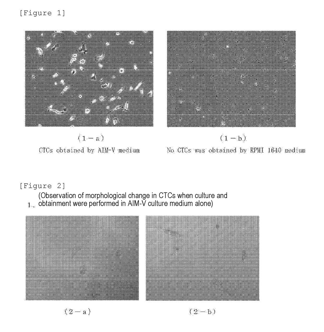
[0120](Test 1: Study on CTCs-Inducing Ability Using RPMI-1640)

[0121]RPMI-1640 was used as a culture medium, and this was used as a common culture medium. The culture medium was divided into a fetal bovine serum (FBS) supplemented group and an autologous serum (Auto Serum) supplemented group. Samples collected from 6 cancer patients were studied for the presence or absence of the CTCs-inducing ability.

[0122]...

example 3

[0153][Selection Test (II) of Efficient Culture Medium for Cancer Cell (CTC) Obtainment—Influence of Additive]

[0154]

[0155]The following culture media were prepared and used as culture media to be tested.

(1) AIM-V (serum-free culture medium)

(2) AS (Auto Serum: autologous serum)

(3) LPS (lipopolysaccharide)

(4) IL-1α (interleukin 1α)

(5) IL-1β (interleukin 1β)

(6) Palmitic Acid

Experimental Method

[0156]Prepared peripheral blood mononuclear cells (PBMCs) of a cancer patient were seeded to each well according to the experimental method described in Example 1 using the culture media described above, to test the influence of an additive in each culture medium on cancer cell (CTC) yield efficiency (cancer cell detection accuracy).

[0157](Test 8: Influence of LPS Addition to AIM-V Culture Medium)

[0158]An AIM-V culture medium (AIM-V: Group A) was used as a basis. A group in which AS (autologous serum) was added thereto (AIM-V+AS: Group B), and further, groups in which LPS (lipopolysaccharide) was ...


PUM


Abstract

A method for detecting or separating/obtaining CTC, which is capable of reliably and stably detecting or separating/obtaining a circulating tumor cell and a circulating tumor stem cell present in trace amounts in a biological circulating fluid such as blood or lymph, even in the state where the cancer type of the tumor cells cannot be determined yet and the state where the tumor cells are present in trace amounts in the biological circulating fluid. The method is attained by detecting or separating/obtaining CTC and/or CTSC in a biological circulating fluid, comprising treatment steps (1) to (4): (1) pretreating a sample from the biological circulating fluid to obtain a mononuclear cell phase; (2) providing a well plate in which a culture medium consisting of a serum-free cell growth medium for circulating tumor cell and/or circulating tumor stem cell has been injected, and seeding thereto the mononuclear cells obtained in step (1), followed by incubation; (3) removing the culture medium from a well of the plate obtained by the incubation in step (2); and (4) detecting or separating/obtaining an adherent tumor cell attached to the well of the plate after step (3).

Description


Claims


Application Information

Patent Timeline
Owner UMEZU YASUIKI
Why Eureka
Try Eureka