Method of evaluating cancer type

a cancer type and cancer technology, applied in the field of cancer type evaluation, can solve the problems of reducing the detection specificity and detection sensitivity of ct (computer tomography), reducing the time, physical and financial burden of the examinee, and reducing the efficiency of examination

Inactive Publication Date: 2011-04-21
AJINOMOTO CO INC
View PDF18 Cites 12 Cited by
  • Summary
  • Abstract
  • Description
  • Claims
  • Application Information

AI Technical Summary

Benefits of technology

[0029]It is an object of the present invention to at least partially solve the problems in the conventional technology. The present invention is made in view of the problem described above, and an object of the present invention is to provide a method of evaluating cancer type, which is capable of evaluating the cancer type accurately by utilizing the concentration of the amino acid related to states of various cancers among amino acids in blood. Specifically, an object thereof is to provide the method of evaluating cancer type, which is capable of narrowing an examinee likely to contract a plura

Problems solved by technology

However, diagnosis based on a fecal occult blood test does not serve as definitive diagnosis, and most of the persons with positive-finding are false-positive.
Furthermore, in regard to early colon cancer, there is a concern that both the detection sensitivity and the detection specificity become lower in the diagnosis based on a fecal occult blood test.
Diagnostic imaging by CT (computer tomography), MRI (magnetic resonance imaging), PET (positron emission computerized-tomography) or the like is not suitable for the diagnosis of colon cancer.
On the other hand, colon biopsy by colonoscopy serves as definitive diagnosis, but is a highly invasive examination, and implementing colon biopsy at the screening stage is not practical.
Furthermore, invasive diagnosis such as colon biopsy gives a burden to patients, such as accompanying pain, and there may also be a risk of bleeding upon examination, or the like.
However, diagnosis by imaging does not serve as definitive diagnosis.
Furthermore, in the case of chest X-ray examination, the detection sensitivity is low, and some examination results according to the Ministry of Health, Labour and Welfare of Japan also report that about 80% of patients who developed lung cancer were overlooked.
Particularly, in early lung cancer, there is a concern that diagnosis by imaging is even poorer in both detection sensitivity and detection specificity.
In chest X-ray examination, there is also a problem of exposure of test subjects to radiation.
Diagnostic imaging by CT, MRI, PET or the like also is not suitable to be carried out as mass screening, from the viewpoints of facilities and costs.
On the other hand, lung biopsy using a bronchoscope, a percutaneous needle, exploratory thoracotomy or a thoracoscope serves as definitive diagnosis, but is a highly invasive examination, and implementing lung biopsy on all patients who are suspected of having lung cancer as a result of diagnostic imaging, is not practical.
Furthermore, such invasive diagnosis gives a burden to patients, such as accompanying pain, and there may also be a risk of bleeding upon examination, or the like.
However, self examination, palpation and visual inspection, and diagnostic imaging do not serve as definitive diagnosis.
In particular, self examination is not effective to the extent of lowering the rate of deaths from breast cancer.
Furthermore, self examination does not enable the discovery of a large number of early cancers, as regular screening by a mammographic examination does.
In early breast cancer, there is a concern that self examination, palpation and visual inspection, or diagnostic imaging is even poorer

Method used


Image

  • Method of evaluating cancer type
  • Method of evaluating cancer type
  • Method of evaluating cancer type

Examples


first embodiment

1-1. Outline of the Invention

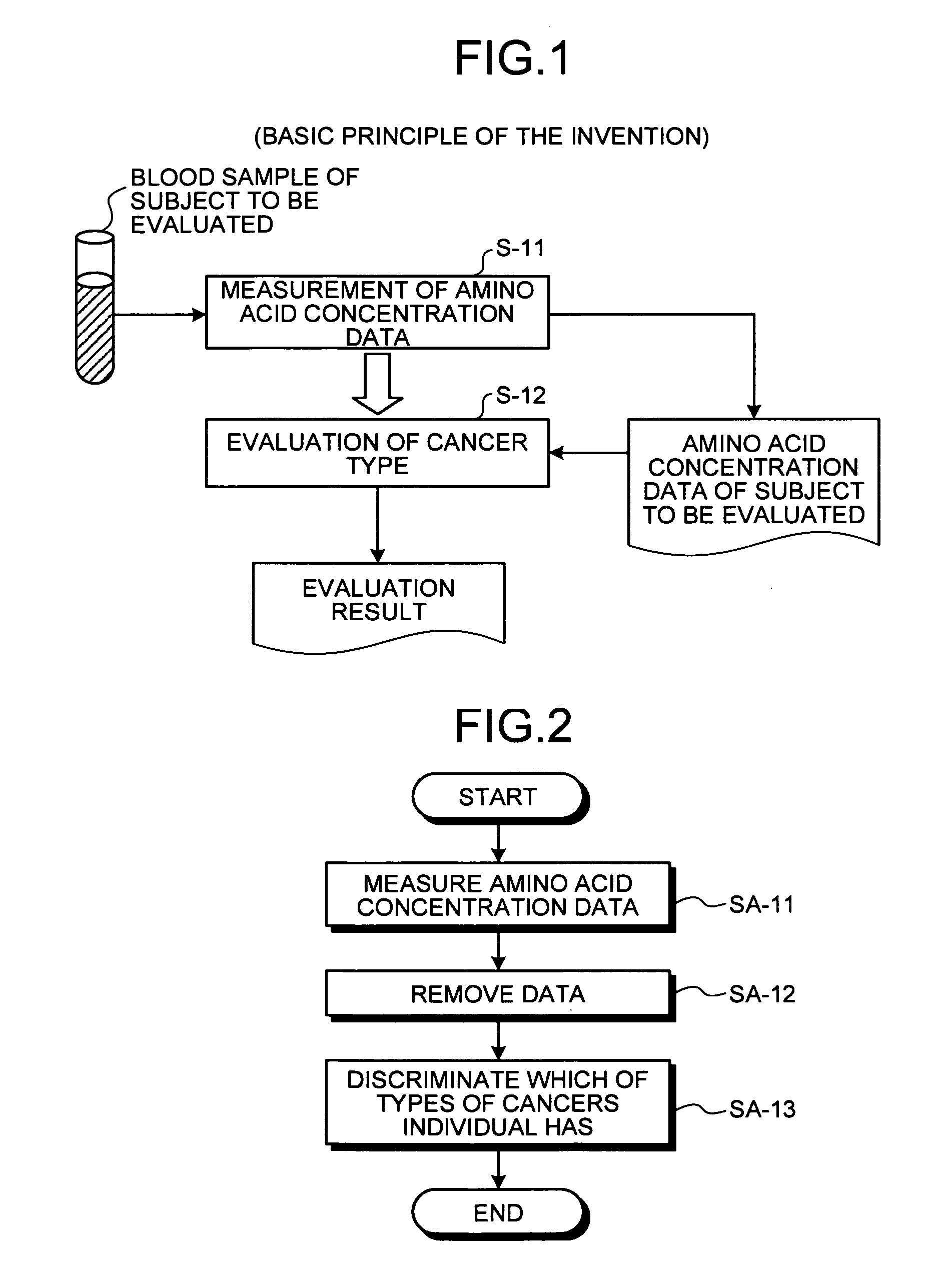
[0219]Here, an outline of the method of evaluating cancer type of the present invention will be described with reference to FIG. 1. FIG. 1 is a principle configurational diagram showing a basic principle of the present invention.

[0220]In the present invention, amino acid concentration data on a concentration value of an amino acid in blood collected from a subject (for example, an individual such as animal or human) to be evaluated is first measured (step S-11). Concentrations of amino acids in blood are analyzed in the following manner. A blood sample is collected in a heparin-treated tube, and then the blood plasma is separated by centrifugation of the collected blood sample. All blood plasma samples separated are frozen and stored at −70° C. before a measurement of amino acid concentrations. Before the measurement of amino acid concentrations, the blood plasma samples are deproteinized by adding sulfosalicylic acid to a concentration of 3%. An amino a...

second embodiment

2-1. Outline of the Invention

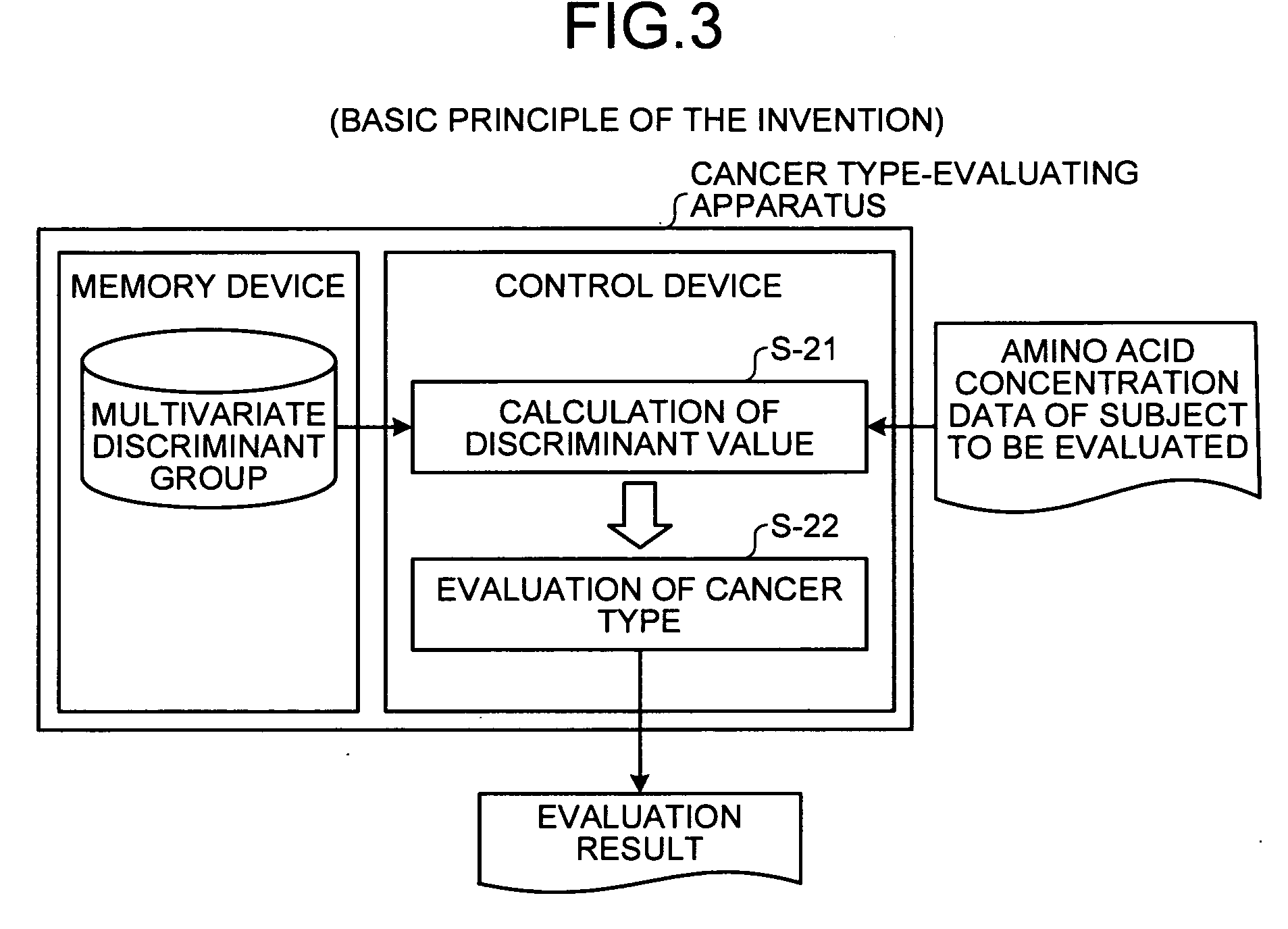
[0271]Herein, an outline of the cancer type-evaluating apparatus, the cancer type-evaluating method, the cancer type-evaluating system, the cancer type-evaluating program and the recording medium of the present invention are described in detail with reference to FIG. 3. FIG. 3 is a principle configurational diagram showing a basic principle of the present invention.

[0272]In the present invention, a discriminant value that is a value of a multivariate discriminant with a concentration of an amino acid as an explanatory variable is calculated in a control device for each of the multivariate discriminants composing a multivariate discriminant group, based on both (a) a concentration value of at least one of Glu, ABA, Val, Met, Pro, Phe, Thr, Ile, Leu, and His contained in previously obtained amino acid concentration data on the concentration value of the amino acid of a subject (for example, an individual such as animal or human) to be evaluated and (b) the...

example 1

[0436]Amino acid concentration in blood is measured by the amino acid analysis method in blood samples of various cancer patient groups with definitive diagnosis of cancer and blood samples of a cancer-free group. The unit of the amino acid concentration is nmol / ml. FIGS. 23 and 24 are boxplots showing the distribution of amino acid explanatory variables of various cancer patients and cancer-free subjects. FIG. 23 is the boxplots showing the distribution of the amino acid explanatory variables of male various cancer patients and male cancer-free subjects, and FIG. 24 is the boxplots showing the distribution of the amino acid explanatory variables of female various cancer patients and female cancer-free subjects. In FIGS. 23 and 24, the horizontal axis indicates the cancer-free group and the various cancer groups, and ABA in the figures represents α-ABA (α-aminobutyric acid). Further, for the purpose of discrimination among the various cancer groups and the cancer-free group, evaluat...


PUM


Abstract

According to the method of evaluating cancer type of the present invention, amino acid concentration data on concentration values of amino acids in blood collected from a subject to be evaluated is measured, and the cancer type in the subject is evaluated based on the concentration value of at least one of Glu, ABA, Val, Met, Pro, Phe, Thr, Ile, Leu, and His contained in the measured amino acid concentration data of the subject.

Description

[0001]This application is a Continuation of PCT / JP2009 / 054091, filed Mar. 4, 2009, which claims priority from Japanese patent application JP 2008-054114 filed Mar. 4, 2008. The contents of each of the aforementioned application are incorporated herein by reference in their entirety.BACKGROUND OF THE INVENTION[0002]1. Field of the Invention[0003]The present invention relates to a method of evaluating cancer type, which utilizes a concentration of an amino acid in blood (plasma).[0004]2. Description of the Related Art[0005]The number of deaths from cancer in Japan in 2004 is 193075 males and 127259 females, and the number of deaths ranks first among the total numbers of deaths. The survival rate may be dependent on the type of cancer, but there are some types for which the five-year survival rate of early stage cancer is 80% or higher, while there are also some types for which the five-year survival rate of progressive cancer is extremely low, such as about 10%. Therefore, early detec...

Claims


Application Information

Patent Timeline
IPC IPC(8): C12Q1/02
CPCG01N33/6806G01N33/574
Inventor IMAIZUMI, AKIRAANDO, TOSHIHIKOOKAMOTO, NAOYUKIIMAMURA, FUMIOHIGASHIYAMA, MASAHIKO
Owner AJINOMOTO CO INC
Why Eureka
Try Eureka