Method for expansion of tumour-reactive T-lymphocytes for immunotherapy of patients with specific cancer types

a tumour-reactive and immunotherapy technology, applied in the field of improved methods for expanding and activating tumour-reactive lymphocytes, can solve the problems of breast cancer that is still poorly understood, disease is usually deadly, and the disease is generally very poor

Active Publication Date: 2011-05-31
SENTOCLONE INT AB
View PDF6 Cites 12 Cited by
  • Summary
  • Abstract
  • Description
  • Claims
  • Application Information

AI Technical Summary

Benefits of technology

[0043]The inventors have found that an expansion method comprising two different phases are especially useful for obtaining a high number of tumour-reactive CD4+ helper and / or CD8+ T-lymphocytes in a relatively short time span, the two phases beingi) a first phase of stimulating tumour-reactive T-lymphocytes with tumour-derived antigen together with at least one substance having agonistic activity towards the IL-2 receptor, to promote survival of tumour-reactive T-lymphocytes, andii) a second phase of activating and promoting growth of tumour-reactive T-lymphocytes, wherein the second phase ii) is initiated when the CD25 cell surface marker (IL-2R marker) is down-regulated on T-lymphocytes.
[0136]Accordingly, the present invention relates to a method for the preparation of memory T-lymphocytes. Normally, when a culture of tumour-reactive T-lymphocytes are expanded according to the present invention from about 35% to about 90% of tumour-reactive T-lymphocytes of the memory type, such as, e.g. from about 40% to about 90%, from about 50% to about 80% or from about 60% to about 70%, will be obtained. The present inventors speculates that the fact that the lymphocytes in phase i) are allowed to regenerated before tumour antigen is added, together with the relatively slow expansion phase leads to formation of a high ratio of memory lymphocytes to effector lymphocytes.

Problems solved by technology

Prognosis of the disease is generally very poor.
If the cancer cannot be removed, the disease is usually deadly within 3 to 6 months.
Despite much research, the cause of breast cancer is still poorly understood.
There are no known effective treatments today, and the prognosis is dismal.
Carcinoid tumors are generally slow-growing.
Radiation therapy and chemotherapy are usually ineffective against leiomyosarcoma.
Thus, the immune system is capable of recognizing and eliminating tumour cells but unfortunately tumours often interfere with the development and function of immune responses.
However, life-threatening toxicity often develops, largely due to the release of tumour necrosis factor (TNF) from activated NK cells.

Method used


Image

  • Method for expansion of tumour-reactive T-lymphocytes for immunotherapy of patients with specific cancer types
  • Method for expansion of tumour-reactive T-lymphocytes for immunotherapy of patients with specific cancer types
  • Method for expansion of tumour-reactive T-lymphocytes for immunotherapy of patients with specific cancer types

Examples


example 1

Expansion of Tumour-Reactive T-Lymphocytes

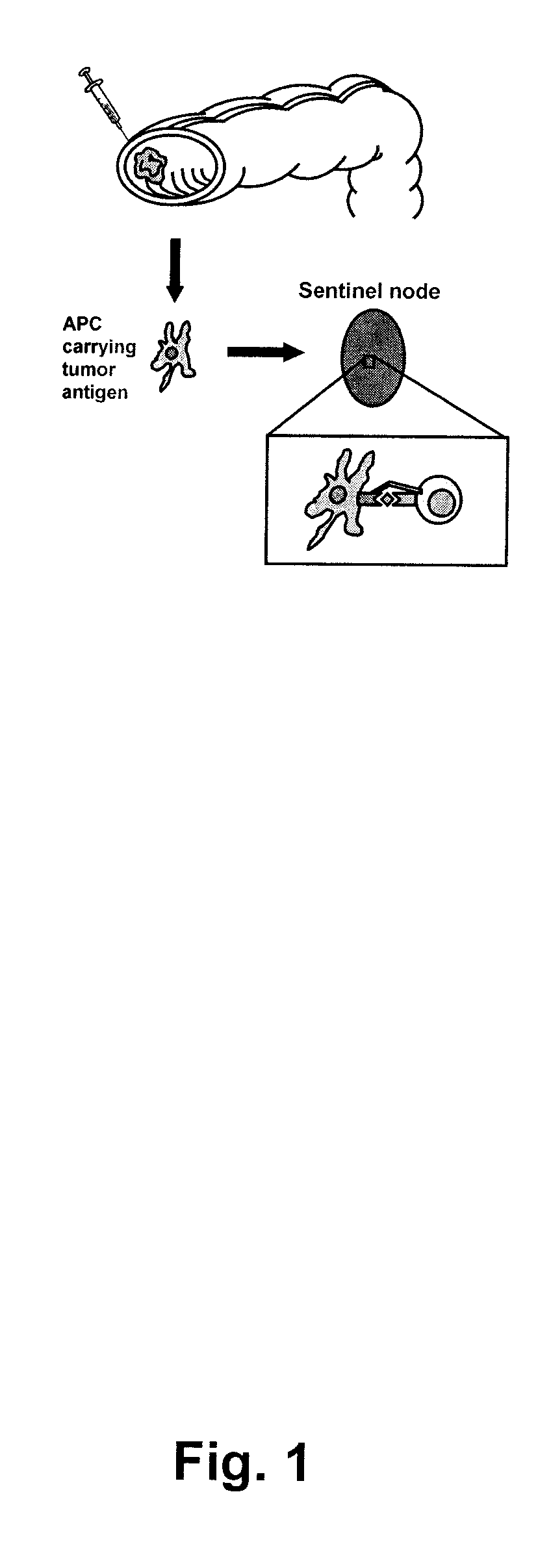
[0174]Identification of sentinel nodes was done peroperatively using the sentinel node technique. Briefly, 1 ml of Patent blue dye was injected (Guerbet, Paris) and distributed superficially in the serosa around the tumour. Within five to ten minutes, one to three mesenteric lymph nodes were coloured blue, these sentinel nodes were marked with sutures and removed (see FIG. 1). One non-sentinel mesenteric lymph node, distant from the tumour, was also identified and removed as a control.

[0175]The sentinel- and non-sentinel lymph nodes were cut in half and 1 mm thick slices were taken from the center and the periphery. The rest of the lymph nodes were sent for histopathological examination according to routine procedure. A part of the tumour, including a sample of the invasive margin, was also removed for research purposes.

Cell Culture

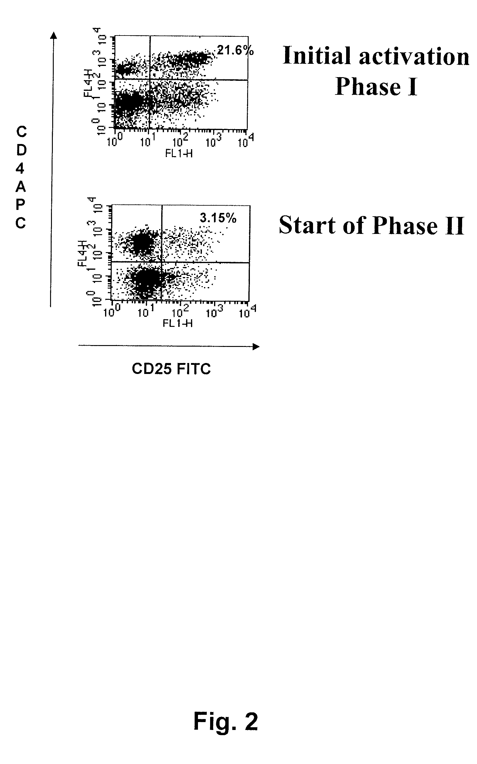
Phase 1, Initial Activation

[0176]The sentinel node material was kept on ice and immediately taken care of using...

example 2

Treatment of Colon Cancer by Administering Tumour-Reactive T-Lymphocytes

Identification and Removal of Sentinel and Metinel Lymph Nodes from Colon Cancer Patients:

[0185]Sixteen patients diagnosed with colon cancer, six woman and ten men with an average age of 62 years were studied. Patients were histopathologically classified as Duke's C or D. There were also 5 patients with Duke's B with aggressive tumour characteristics such as ulcerations, vascular or perineural invasion. Patients 7 and 14 however had earlier been surgically treated due to colon cancer and now had recurrent disease with metastases to the liver. The local ethical committee approved the study and each patient gave informed consent.

[0186]Identification of sentinel or metinel nodes was done intraoperatively. Mobilisation of the colonic tumour site was achieved by division of peritoneal adhesions in order to facilitate inspection of tumour and mesentery. Injections of Patent blue dye (Guerbet, Paris) were distributed s...

example 3

Expansion of Lymph Node Acquired Lymphocytes from Patient with Gallbladder Cancer

[0203]On day one, cell cultures containing approximately 35 million (Culture 1) and 5 million cells (Culture 2) was set up.

[0204]Throughout the whole culturing period, cells were kept in incubator at 37° C. and 5% CO2 and 240 IU IL-2 / mL media added every 3-4 days.

[0205]Initially, both cultures contained a high proportion of red blood cells which eventually decreased as only lymphocytes proliferated in the cultures.

[0206]On day 3, autologous tumor extract were added to both cultures at a concentration of 1 / 100.

[0207]On day 10, phase II was initiated in both cultures by adding radiated antigen presenting cells, autologous tumor extract and interleukin-2 (240 IU / ml) to the cultures. At this stage, the cultures contained approximately 45 million and 15 million lymphocytes, respectively.

[0208]Cultures were maintained regularly by keeping the cell concentration at approximately 3 million cells / ml.

[0209]Cells ...


PUM

PropertyMeasurementUnit
timeaaaaaaaaaa
timeaaaaaaaaaa
timeaaaaaaaaaa

Abstract

Methods for treating a patient suffering from a neoplastic disease are disclosed and described. A number of diseases can be treated under the present methods, including without limitation gall bladder cancer, hepato cellular cancer, ovarian cancer, small intestine cancer, lung cancer, mesothelioma, breast cancer, kidney cancer, pancreas cancer, prostate cancer, carcinoid cancer, leiomyosarcoma, or metastasis thereof. A general method for providing such treatment may include: 1) identifying in a patient one or more sentinel and / or metinel lymph nodes draining the neoplasm; 2) resecting the one or more nodes and, optionally all or part of the tumour or metastasis; 3) isolating tumour-reactive T-lymphocytes from said lymph nodes; 4) in vitro expanding said tumour-reactive T-lymphocytes; and 5) administering the thus obtained tumour-reactive T-lymphocytes to the patient.

Description

PRIORITY CLAIM[0001]This application claims the benefit of earlier filed U.S. Provisional Application No. 60 / 937,484, filed Jun. 27, 2007, which is incorporated herein by reference.FIELD OF THE INVENTION[0002]The invention relates to an improved method for expansion and activation of tumour-reactive lymphocytes, in particular CD4+ helper and / or CD8+ T-lymphocytes. The T-lymphocytes are not CD4+ CD25+Hi lymphocytes, i.e. the present invention does not cover regulatory T-lymphocytes. The lymphocytes may be used for treating and / or preventing specific cancer types, in particular gall bladder cancer, hepato cellular cancer, ovarian cancer, small intestine cancer, lung cancer, mesothelioma, breast cancer, kidney cancer, pancreas cancer, prostate cancer, carcinoid cancer, leiomyosarcoma, or metastasis thereof.BACKGROUND OF THE INVENTION[0003]According to the immune surveillance hypothesis, the immune system is continuously sensitized against developing tumours, where experimental evidence...

Claims


Application Information

Patent Timeline
Patent Type & Authority Patents(United States)
IPC IPC(8): A01N63/02A61K35/26A61K35/02A61K39/00
CPCA61K39/0011A61K2039/5158
Inventor WINQVIST, OLATHORN, MAGNUS
Owner SENTOCLONE INT AB
Why Eureka
Try Eureka