Differential Identification of Pancreatic Cysts

a technology of pancreatic cysts and differential identification, applied in the field of diagnosis, can solve the problems of difficult to determine the type of cyst from conventional clinical, radiographic, or cytologic findings, and the management of these cysts is concomitantly becoming a major problem

Inactive Publication Date: 2019-08-22
THE JOHN HOPKINS UNIV SCHOOL OF MEDICINE
View PDF0 Cites 0 Cited by
  • Summary
  • Abstract
  • Description
  • Claims
  • Application Information

AI Technical Summary

Benefits of technology

The invention provides methods and tools to differentially diagnose pancreatic cysts based on the mutations in certain genes. This method can identify the type of pancreatic cyst, such as serous cystadenoma or mucinous cystic neoplasm, and aid in managing medical treatment. The methods involve testing the nucleic acids isolated from cyst fluid or epithelial cells and detecting mutations in genes like VHL, GNAS, RNF43, KRAS, and CTNNB1. A device or kit can also be used for this purpose. Overall, the invention helps in better understanding and managing pancreatic cysts.

Problems solved by technology

Management of these cysts is concomitantly becoming a major clinical problem (1, 2).
But once a cyst is identified, it poses a challenging, life-long management problem (1, 2, 9-13).
Unfortunately, it is often difficult to determine the type of cyst from conventional clinical, radiographic, or cytologic findings (1, 2, 9-16, 17)
The pre-operative diagnosis of surgically-excised cysts has been shown to be erroneous in one third of cases, which can lead to unnecessary surgical procedures (22).
These fluids are often acellular and therefore not typically useful for cytologic diagnosis.

Method used


Image

  • Differential Identification of Pancreatic Cysts
  • Differential Identification of Pancreatic Cysts
  • Differential Identification of Pancreatic Cysts

Examples


example 1

Materials and Methods

Patients and Specimens

[0048]The present study was approved by the Institutional Review Boards of Johns Hopkins Medical Institutions, Memorial Sloan Kettering Cancer Center, Wayne State University Emory University and the University of Indiana. Lesions were classified as IPMNs, MCNs, SPNs or SCAs using standard criteria (74). None of the patients with SCAs had clinical features of the VHL syndrome and all of the SPNs were Stage 1B, pT2N0M0 (74). IPMNs were subtyped by internationally accepted criteria (75).

[0049]Fresh-frozen tissue specimens of surgically resected cystic neoplasms of the pancreas were obtained through the prospectively maintained Johns Hopkins Surgical Pathology Tumor Bank and from collaborating institutions (Memorial Sloan Kettering Cancer Center, Emory University, the University of Utrecht and Wayne State University). IPMNs, SCAs, SPNs and two MCNs specimens were microdissected using a razor blade. In these cases, serial frozen sections were us...

example 2

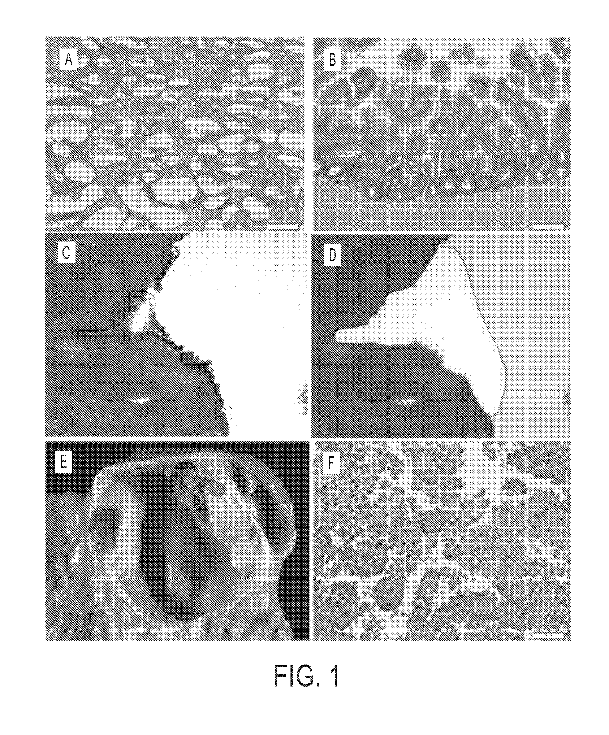
[0061]Experimental design. Neoplastic cysts are composed of a mixture of neoplastic epithelial cells and non-neoplastic cells (stromal, vascular, and inflammatory (17)) (FIG. 1). To maximize our ability to detect mutations, we carefully microdissected the neoplastic epithelial cells from the non-neoplastic cells. This was most difficult in the MCNs because of the cellular ovarian-type stroma present in these lesions (FIG. 1C, D). Following microdissection, the neoplastic cell content of each of the cyst samples analyzed in this study was at least 33%.

[0062]DNA from 32 microdissected cysts, eight of each of the four types, as well as matched DNA from normal tissues of the same patients, was used in this study. The clinical and histopathologic characteristics of the patients and their cystic lesions are detailed in Table S 1. The DNA was ligated to adapters and amplified using standard Illumina protocols. The amplified DNA was then captured with a 50 MB SureSelect Enrichment System. T...

example 3

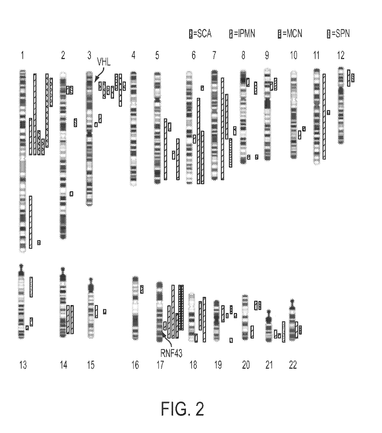
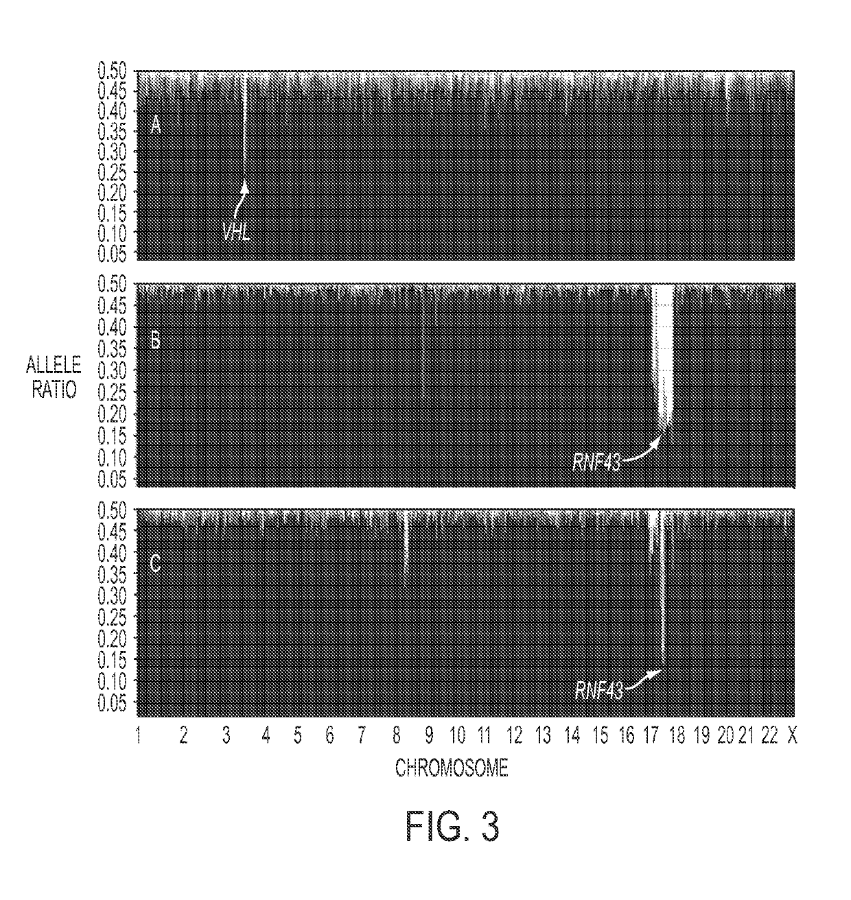
[0063]Analysis of SCAs. Using single nucleotide polymorphisms in the sequences captured by the SureSelect Enrichment System, we were able to identify 15,190±428 heterozygous variants in the matched normal DNA samples of the eight SCA patients. Loss of heterozygosity (LOH) of at least one chromosomal region was identified in each of the eight SCAs studied (FIG. 2, Table S3). The maximum degree of LOH (70%±13%) confirmed the high fraction of neoplastic cell content achieved upon microdissection. The only region that was lost in the majority of SCAs was on chromosome 3p (FIG. 2, example in FIG. 3A). Seven of the eight SCAs lost chromosome 3p alleles, with the losses demarcated by bases 9,934,713 to 12,850,443. To determine whether this LOH was associated with reduplication of the remaining allele, we compared the copy number of all sequences lying within the regions of LOH to those of all other chromosomal regions in the same cysts. This was accomplished by comparing normalized tag cou...


PUM


Abstract

More than 2% of adults harbor a pancreatic cyst, a subset of which progress to invasive lesions with lethal consequences. To assess the genomic landscapes of neoplastic cysts of the pancreas, we determined the exomic sequences of DNA from the neoplastic epithelium of eight surgically resected cysts of each of the major neoplastic cyst types: serous cystadenomas (SCAs), intraductal papillary mucinous neoplasms (IPMNs), mucinous cystic neoplasms (MCNs) and solid pseudo-papillary neoplasms (SPNs). SPNs are low-grade malignancies, and IPMNs and MCNs, but not SCAs, have the capacity to progress to cancer. We found that SCAs, IPMNs, MCNs, and SPNs contained 10=4.6, 27=12, 16=7.6, and 2.9=2.1 somatic mutations per tumor, respectively. Among the mutations identified, E3 ubiquitin ligase components were of particular note. Four of the eight SCAs contained mutations of VHL, a key component of the VHL ubiquitin ligase complex that has previously been associated both with renal cell carcinomas, SCAs, and other neoplasms. Six of the eight IPMNs and three of the eight MCNs harbored mutations of RNF43, a gene coding for a protein with intrinsic E3 ubiquitin ligase activity that has not previously been found to be genetically altered in any human cancer. The preponderance of inactivating mutations in RNF43 unequivocally establish it as a suppressor of both IPMNs and MCNs. SPNs contained remarkably few genetic alterations, but always contained mutations of CTNNB1, previously demonstrated to inhibit degradation of the encoded protein (β-catenin) by E3 ubiquitin ligases. These results highlight the essential role of ubiquitin ligases in these neoplasms and have important implications for the diagnosis and treatment of patients with cystic tumors.

Description

[0001]The invention was made with government support under CA 62924, CA 57345, and CA 43460 awarded by National Institutes of Health. The government has certain rights in the invention.TECHNICAL FIELD OF THE INVENTION[0002]This invention is related to the area of cancer management. In particular, it relates to the areas of prognosis and diagnosis.BACKGROUND OF THE INVENTION[0003]As the result of the increasing use of abdominal imaging in standard medical practice, pancreatic cysts are being identified with increasing frequency. Management of these cysts is concomitantly becoming a major clinical problem (1, 2). These lesions occur in more than 20% of patients examined at autopsy (3), in as many as 19.6% of patients evaluated by MRI (4-6), and as many as 2.6% of patients evaluated by CT (7, 8). In the vast majority of cases, the cysts are identified as incidental findings in patients undergoing imaging for symptoms unrelated to pancreatic pathology. But once a cyst is identified, it ...

Claims


Application Information

Patent Timeline
Patent Type & Authority Applications(United States)
IPC IPC(8): C12Q1/6886C12Q1/6883
CPCC12Q1/6886C12Q1/6883C12Q2600/156
Inventor VOGELSTEIN, BERTKINZLER, KENNETH W.PAPADOPOULOS, NICKOLASWU, JIANHRUBAN, RALPHMAITRA, ANIRBANDAL MOLIN, MARCO
Owner THE JOHN HOPKINS UNIV SCHOOL OF MEDICINE
Why Eureka
Try Eureka