Monitoring a respiratory curve

a technology of respiratory curve and current respiratory curve, which is applied in the field of monitoring a current respiratory curve of a patient, can solve the problems of disadvantageous dependence of signal quality and substantial inability to use motion sensors

Active Publication Date: 2020-03-12
SIEMENS HEALTHCARE GMBH
View PDF2 Cites 4 Cited by
  • Summary
  • Abstract
  • Description
  • Claims
  • Application Information

AI Technical Summary

Benefits of technology

[0052]The performance of the disclosure in the form of a computer program or a computer-readable data carrier including program code of a computer program offers t

Problems solved by technology

The motion sensors, however, are arranged in the immediate vicinity of or directly on the body of the patient, with the result that their signal quality is disadvantageously dependent on the positioning of the sensor or the physiology/positioning of the patient.
For particularly large/small patients, motion sensors

Method used


Image

  • Monitoring a respiratory curve
  • Monitoring a respiratory curve
  • Monitoring a respiratory curve

Examples


Embodiment Construction

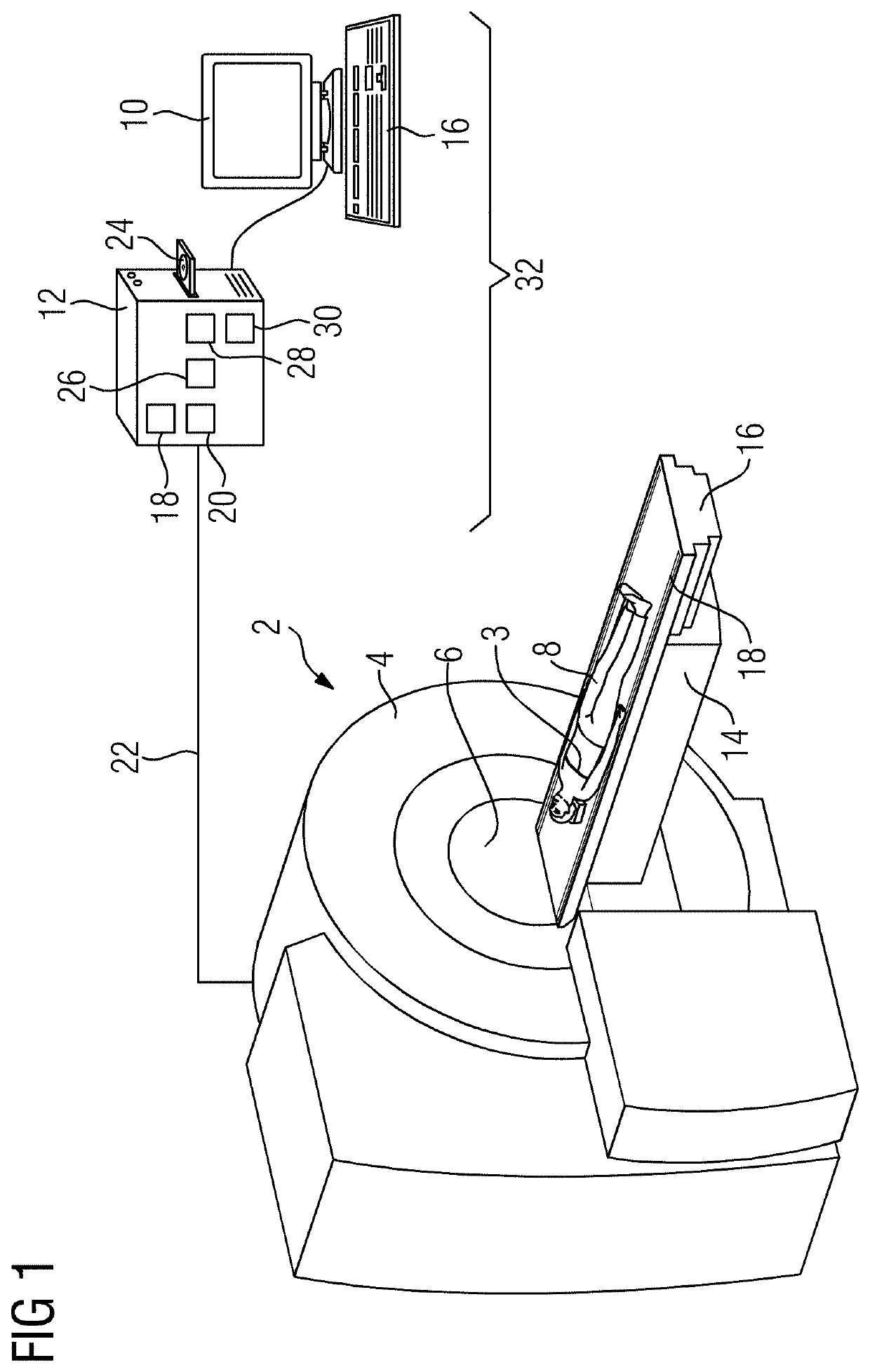
[0059]The medical imaging system 2 shown in FIG. 1 in the form of a magnetic resonance system includes a hollow cylindrical-shaped base unit 4 in the interior of which during operation, the so-called tunnel 6, an electromagnetic field is generated for a magnetic resonance scanning or investigation of an examination object in the form of a patient 8. Additionally, a patient table 14 with a movable support board 16 is provided. The patient 8 may be positioned, as shown, on the support board 16. The patient table 14 is positioned outside the base unit 4 so that the support board 16 together with the patient 8 may be moved at least partially into the tunnel 6 for the examination. In this embodiment, a sensor unit 3 in the form of a chest belt is attached to the patient 8 at chest height. The chest belt 3 serves to acquire a respiratory curve A of the patient 8, in particular a reference respiratory curve REFA and a current respiratory curve AKA.

[0060]The tomograph 2 has a computation un...


PUM


Abstract

A method is provided for monitoring a current respiratory curve of a patient with regard to a recording region which is imaged by magnetic resonance scanning. The method includes acquiring a reference respiratory curve of the patient over a plurality of respiratory cycles; establishing a respiration state of the patient that is suitable for the magnetic resonance scanning based on the reference respiratory curve; determining at least one reference recording time window and a trigger threshold value for starting a magnetic resonance scan based on the previously determined respiration state; carrying out at least one magnetic resonance scan within the determined reference recording time window of the current respiratory curve using the trigger threshold value; and continually acquiring and monitoring the current respiratory curve during the magnetic resonance scan in the reference recording time window.

Description

[0001]The present patent document claims the benefit of German Patent Application No. 10 2018 215 415.5, filed Sep. 11, 2018, which is hereby incorporated by reference.TECHNICAL FIELD[0002]The disclosure relates to monitoring a current respiratory curve of a patient.BACKGROUND[0003]Magnetic resonance imaging is a widespread technology for generating magnetic resonance images of a body interior of a patient. It relies on the physical phenomenon of nuclear magnetic resonance (MR). It is distinguished by large and variable soft tissue contrasts. For this purpose, with the aid of an MR apparatus, excitation pulses of a pulse sequence are radiated into the patient and initiate MR signals in the patient. The MR signals may be received by MR coils and are passed on to an evaluating unit in order to generate an MR image of the patient from the MR signals. If magnetic resonance data of a patient is recorded in the region of the torso, (e.g., the thoracic and / or abdominal regions), the corres...

Claims


Application Information

Patent Timeline
IPC IPC(8): A61B5/00A61B5/055A61B5/113
CPCA61B5/113A61B5/7292A61B5/055A61B5/742A61B5/7285A61B5/7289G01R33/5673
Inventor KARTÄUSCH, RALFPAUL, DOMINIK
Owner SIEMENS HEALTHCARE GMBH
Why Eureka
Try Eureka