Automatic Detection and Quantification of the Aorta from Medical Images

a technology for aortic diseases and quantification, applied in the field of detection and quantification of aortic diseases of patients from medical images, can solve the problems of delayed diagnosis or failure to diagnose, severe and potentially fatal consequences of aortic diseases, and asymptomatic aortic diseases

Active Publication Date: 2020-05-21
SIEMENS HEALTHCARE GMBH
View PDF4 Cites 8 Cited by
  • Summary
  • Abstract
  • Description
  • Claims
  • Application Information

AI Technical Summary

Benefits of technology

[0003]In accordance with one or more embodiments, systems and methods are provided for evaluating an aorta of a patient. A medical image of an aorta of a patient is received and the aorta is segmented from the medical image. A measurement plane is identified on the segmented aorta and a measurement is calculated at the measurement plane. The aorta of the patient is evaluated based on the measurement cal

Problems solved by technology

Left untreated, such aortic diseases have severe and potentially fatal consequences.
However, such aortic diseases are often asymptomatic and are only detected after complicati

Method used


Image

  • Automatic Detection and Quantification of the Aorta from Medical Images
  • Automatic Detection and Quantification of the Aorta from Medical Images
  • Automatic Detection and Quantification of the Aorta from Medical Images

Examples


Embodiment Construction

[0017]The present invention generally relates to methods and systems for the automatic detection and quantification of the aorta from medical images. Embodiments of the present invention are described herein to give a visual understanding of such methods and systems. A digital image is often composed of digital representations of one or more objects (or shapes). The digital representation of an object is often described herein in terms of identifying and manipulating the objects. Such manipulations are virtual manipulations accomplished in the memory or other circuitry / hardware of a computer system. Accordingly, is to be understood that embodiments of the present invention may be performed by a computer system using data stored within the computer system.

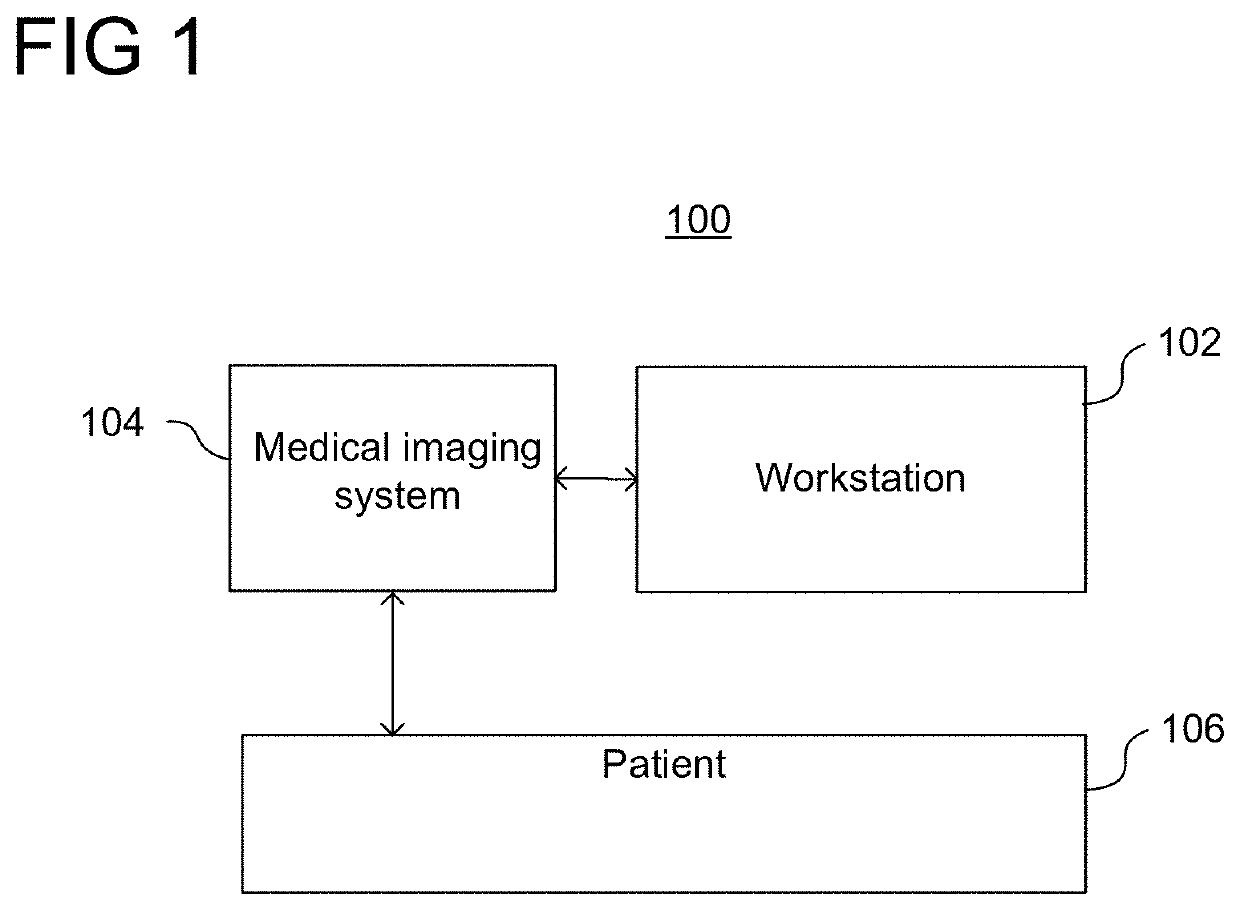
[0018]FIG. 1 shows a system 100 configured to evaluate an aorta of a patient, in accordance with one or more embodiments. System 100 includes workstation 102, which may be used for assisting a clinician (e.g., a doctor, a medical pr...


PUM


Abstract

Systems and methods are provided for evaluating an aorta of a patient. A medical image of an aorta of a patient is received. The aorta is segmented from the medical image. One or more measurement planes are identified on the segmented aorta. At least one measurement is calculated at each of the one or more measurement planes. The aorta of the patient is evaluated based on the at least one measurement calculated at each of the one or more measurement planes.

Description

TECHNICAL FIELD[0001]The present invention relates generally to detection and quantification of an aorta of a patient from medical images, and more particularly to the automatic detection and quantification of the aorta using a plurality of machine learning models to analyze medical images.BACKGROUND[0002]Early detection is critical for many aortic diseases, such as aortic dissection, aortic rupture, and ruptured abdominal aortic aneurysms. Left untreated, such aortic diseases have severe and potentially fatal consequences. Conventionally, such aortic diseases are diagnosed by manual evaluation of medical images of a patient. However, such aortic diseases are often asymptomatic and are only detected after complications have manifested. It is therefore difficult to diagnose such aortic diseases before complications have manifested, resulting in a delayed diagnosis or a failure to diagnose.BRIEF SUMMARY OF THE INVENTION[0003]In accordance with one or more embodiments, systems and meth...

Claims


Application Information

Patent Timeline
IPC IPC(8): G06T7/11G06K9/46G06T7/00G16H30/40
CPCG16H30/40G06T7/0012G06T2200/04G06T2207/30101G06T2207/10081G06T7/12G06K9/4638G06K2209/051G06T7/11G06T7/62G06N20/00G06V2201/031
Inventor RAPAKA, SAIKIRANGULSUN, MEHMET AKIFNEUMANN, DOMINIKSPERL, JONATHANKAERGEL, RAINERGEORGESCU, BOGDANSHARMA, PUNEET
Owner SIEMENS HEALTHCARE GMBH
Why Eureka
Try Eureka