Apparatus and method for contrast-enhanced ultrasound imaging
a technology of contrast enhancement and ultrasound, applied in image enhancement, instruments, ultrasonic/sonic/infrasonic image/data processing, etc., can solve the problems of hardly achievable exact relationship between the acquired ultrasound signal and the local microbubble concentration, and the difficulty of quantification, etc., to maximize the inter-correlation coefficient
Active Publication Date: 2020-06-25
INST NAT DE LA SANTE & DE LA RECHERCHE MEDICALE (INSERM) +4
View PDF1 Cites 0 Cited by
- Summary
- Abstract
- Description
- Claims
- Application Information
AI Technical Summary
Benefits of technology
[0030]More specifically, the step of compensating said relative movements may comprise maximizing a
Problems solved by technology
However, using the TIC-based approach, the quantification is typically hampered by two main reasons: first, the exac
Method used
View moreImage
Smart ImageViewing Examples

Examples
 PUM
 Abstract
An apparatus and a method for contrast-enhanced ultrasound (CEUS) including use of a fluid dynamics model for the analysis of dynamic contrast-enhanced ultrasound (DCEUS).
Description
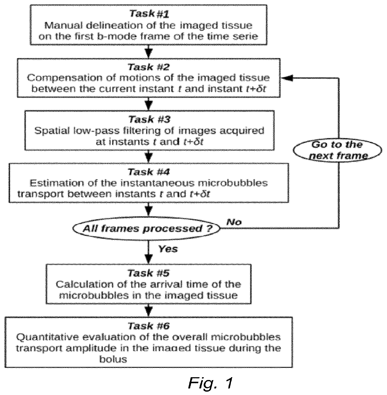
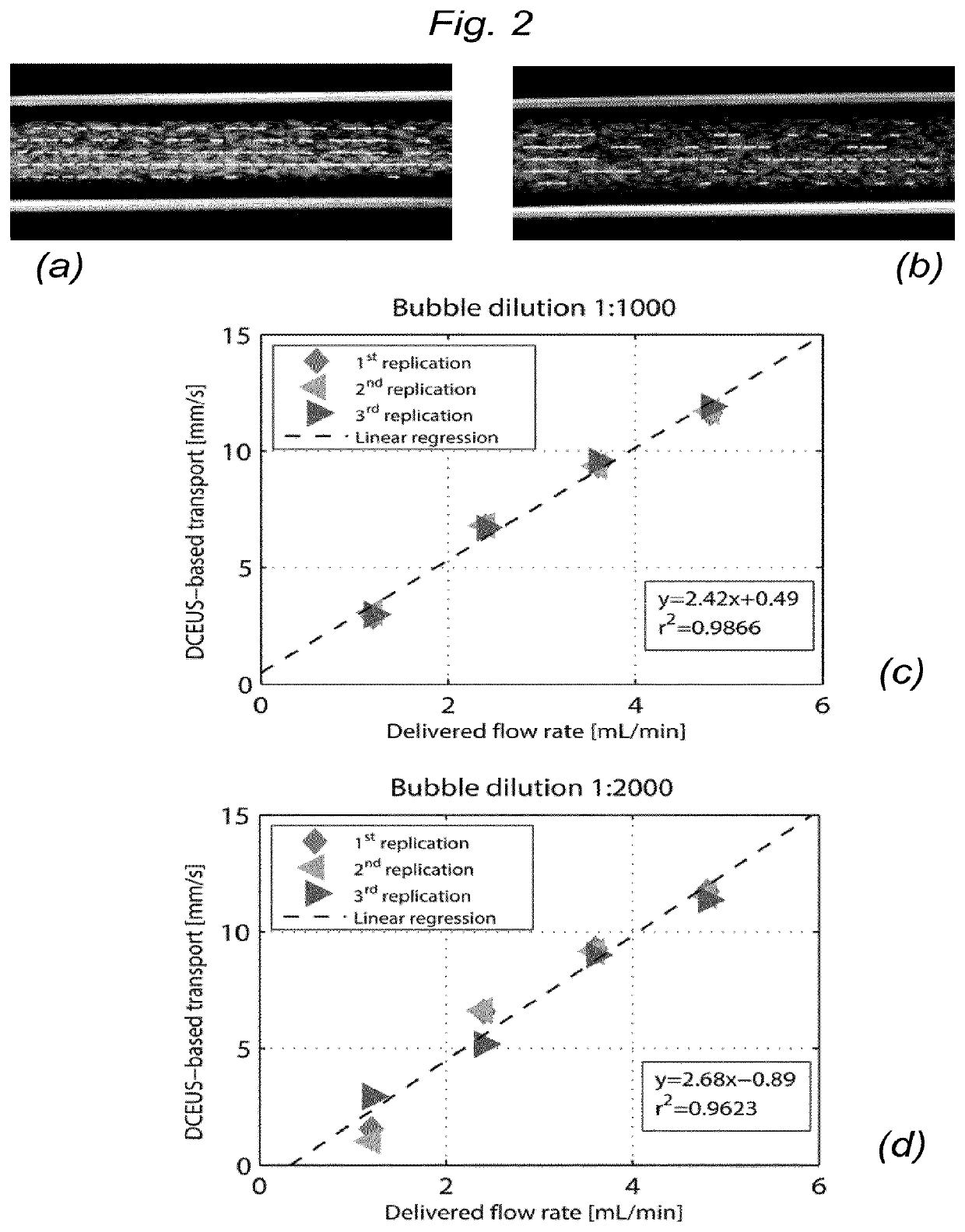
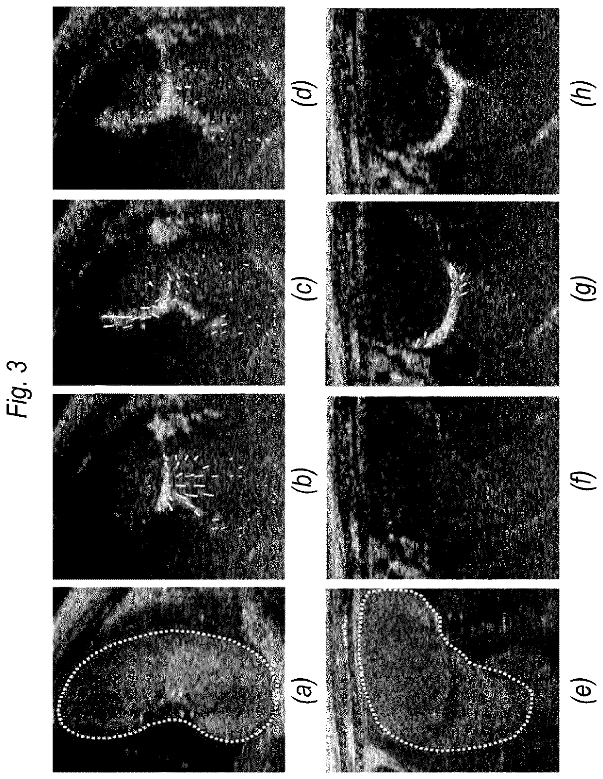
TECHNICAL FIELD[0001]The present disclosure relates to contrast-enhanced ultrasound, also referred to in the abbreviated form as “CEUS”.BACKGROUND[0002]Contrast-enhanced ultrasound is a non-invasive imaging technique that has been used extensively in blood perfusion imaging of various organs. This modality is based on the acoustic detection of gas-filled microbubble contrast agents used as intravascular flow tracers. These contrast agents significantly increase the ultrasound scattering of blood, thus allowing the visualization and the assessment of microcirculation (i.e., blood velocity, blood volume fractions) commonly undetectable by Doppler ultrasound (DUS). The rheology of microbubbles in the microcirculation is similar to that of red blood cells, thus demonstrating the microbubbles do not interfere with hemodynamics and have a good safety profile in both cardiac and abdominal ultrasound applications.[0003]To achieve this objective, computer tools are required for the spatio-te...
Claims
Login to View More Application Information
Patent Timeline
 IPC IPC(8): A61B8/08G06T7/00
CPCG06T2207/10016A61B8/0866A61B8/481A61B8/5276G06T2207/20024G06T2207/10132G06T2207/30104G06T7/0016G01S7/52039
Inventor DENIS DE SENNEVILLE, BAUDOUINPERROTIN, FRANCKESCOFFRE, JEAN-MICHELBOUAKAZ, AYACHE
Owner INST NAT DE LA SANTE & DE LA RECHERCHE MEDICALE (INSERM)
Features
- R&D
- Intellectual Property
- Life Sciences
- Materials
- Tech Scout
- Unparalleled Data Quality
- Higher Quality Content
- 60% Fewer Hallucinations
Learn More Browse by: Latest US Patents, China's latest patents, Technical Efficacy Thesaurus, Application Domain, Technology Topic, Popular Technical Reports.
© 2025 PatSnap. All rights reserved.Legal|Privacy policy|Modern Slavery Act Transparency Statement|Sitemap|About US| Contact US: help@patsnap.com