Intraosseous drilling device with barrel having internal stylet/motor housing with barrel opening extender

a technology of intraosseous drilling and barrel, which is applied in the field of portable and passive safety intraosseous drilling devices, can solve the problems of prone to contact with surfaces at a distance, and achieve the effects of avoiding potential exposure, facilitating manual operation, and simplifying disposal of the sam

Inactive Publication Date: 2020-09-24
JUNE MEDICAL IP LLC
View PDF0 Cites 0 Cited by
  • Summary
  • Abstract
  • Description
  • Claims
  • Application Information

AI Technical Summary

Benefits of technology

[0010]A distinct advantage of the versatile apparatus now disclosed is the provision of a drill with stylet / cannula implement that exhibits total passive safety to avoid any potential exposure to contaminated components subsequent to application of the cannula within a target patient's bone. Another advantage includes the ability to provide a potentially disposable drill / stylet combination that accords the ability to have the entirety of the device provided as a single portable structure, thus eliminating the need to locate separate component parts prior to utilization and simplified disposal of the same. Another advantage is the ability to provide a drill component that has a proper shape that allows for facilitated manual operation for stylet / cannula introduction if needed. Yet another advantage is the ability of the stylet to be retracted back within the drill subsequent to drilling completion and detachment from the cannula. Still another advantage of the overall device and system is the potential for a sensor to determine the exact moment of entry within the intermedullary space of a subject bone, thus eliminating the possibility of such a device from entering such a space too far or not far enough, maximizing the amount of space such a device permits fluid introduction and / or removal from the subject bone. Another advantage is the ability to store the device without any electrical contacts and thus the ability to conserve battery power until utilized, with the potential, as well, to remove any such batteries easily after use for disposal thereof and placement of the remaining device portions within a proper disposal container associated with bodily fluid, and the like, contacted articles. Yet another advantage is the ability to manually activate the device for electrical connection and subsequent movement of an internally stored permanently attached stylet and combined removable needle. Still another advantage is the provision of a recessed hub on the needle with an recessed lock between the drill-attached stylet and needle. Yet another advantage is the facilitation of utilization from transport to disposal, including easily transporting such a device in a pocket or bag, removing it in total, unwrapping from a sealed package, flipping open the device with one hand, manually moving the motor / stylet / cannula sled for external access, operating the drill component to introduce the style / cannula within a subject patient's bone, upon completion, turning the device a quarter (or other degree) turn dislodging and retracting the motor / stylet back into the device with an automatic cover over the stylet point, subsequently dismantling the device for placement in a sharps container and otherwise disposal of the remainder. Still another advantage of a potential embodiment is the ability to provide a light within the device for visibility purposes as well as a possible indicator of proper electrical connections upon engagement between the handle and barrel. Still another advantage is a potential embodiment wherein the handle portion of the device is configured to stand upright on its own and with the barrel attached and manipulated to any angle of rotation around the handle / barrel connector, and the ability of the light (or lights) to remain in operation as the device stands on its own and with the barrel rotated to different angles. Yet another advantage is the potential embodiment of the provision of a protective cap within the barrel over the motor / stylet / cannula that also functions as a extension externally to allow for sufficient space for a user to effectively grasp the cannula hub as it has penetrated a subject patient's skin and bone, and that remains extended subsequent to motor / stylet retraction as a manner of preventing full closure of the device handle back to its original stored position. Still another advantage is a potential embodiment including a protective cap with an extending tab that correlates with openings within the barrel of the device to both serve as an indicator of location of the cap (prior to and after device utilization) as well as a detent to prevent retraction of the cap upon activation of the retraction operation for the motor / stylet.
[0011]Accordingly, the inventive intraosseous device comprises a disposable drill / stylet combination, wherein said drill includes a stylet retraction port therein, a retracting mechanism attached to said stylet and disposed within said drill, and a cannula connected in detachable relation to said stylet, wherein said stylet is permanently attached to said drill, wherein said retracting mechanism operates subsequent to utilization of said drill to introduce said stylet and cannula within the intermedullary space of a subject bone, said retracting mechanism moving said stylet from an external location to said stylet retraction port within said drill subsequent to a drilling operation, and wherein said cannula is attached to said drill with a Luer lock component with a lip portion present external to said drill and an automatic closure internally within said drill that activates upon disengagement of said cannula from said drill. The external lip of the cannula also provides, if needed, a surface for a user to grab while maneuvering the drill. Also encompassed herein is the inclusion of a sensor on said stylet, wherein said sensor measures pressure subsequent to bone entry such that once passed through to said intermedullary space the pressure difference deactivates the drill and activates the retraction mechanism and detaching from said cannula. The method of utilization of such a device (with or without the sensor as described) is also encompassed herein wherein the cannula as retained within the bone and skin and accessing said intermedullary space of the subject bone allows for introduction or removal of fluid, etc., therein.
[0012]Alternatively, the overall device may be provided with an internal movable combination of a stylet and cannula that are not directly connected, but are co-connected through a rotatable base. In such a configuration, the stylet may, for instance, be integrated within such a base in that it may be permanently affixed thereto and aligned with the opening shaft of the cannula so as to egress out of the top with the tapered end available for drill purposes. The cannula may then be attached to such a base through an two or more outer arm extensions from the base that include flanges that insert and turn within complementary structures on the base of the cannula in order to allow simultaneously for temporary attachment (and detachment through turning of the internal drill device base a specific distance after drilling has been completed) and rotation of the cannula in concert with the stylet. The movable nature allows for a stationed motor and gear box within the housing of the drill device with the extendible stylet / cannula combination arranged to retain gear association with the motor and gear box (to permit such rotation for drilling results) while allowing as well for retraction of the stylet once drilling has been effectuated. In addition, an internal seal (or door) may be provided initially to ensure the internal stylet and cannula are protected from contamination prior to use; if so, such a door may be configured to move and thus open to provide an egress point through the distal end of the drill device for the stylet / cannula combination to exit for drilling to commence. At the same time, an opposing door / seal component may be primed upon such extension in order to automatically close upon retraction of the stylet after use and / or upon detachment of the cannula from the internal extendible base (and thus from the drill device itself). In this manner, the passive safety necessity is provided in an alternative manner but still as effective, if not more effective, than the prior disclosure above. The user merely activates the internal base extension to open the drill egress point for the stylet / cannula to exit, activate the motor to effectuate the gear box and extended gear shaft to rotate the stylet and cannula simultaneously for drilling within a target patient's skin and bone until entering the target intermedullary space thereof, stopping such a drilling action, rotating the drill a slight turn to disengage the cannula from the base, at which point the stylet and internal base return automatically within the drill device and the primed door / seal closes automatically as well, leaving only the cannula with its Luer lock end available for attachment with an IV line or like implement as needed for fluid, etc., introduction or material, etc., removal from the target intermedullary bone space. If further desired, the user may include the application of a disinfectant on the Luer lock tip to further ensure contamination is not an issue.
[0013]The disclosed device may alternatively include a cannula with a hub for IV or other connection / insertion (again, for medicament, drug, fluid, etc., delivery within a subject patient's bone) that itself utilizes a novel recessed attachment point, rather than an extended Luer lock tip. Such a recessed configuration would allow for stylet temporary engagement during operation of the intraosseous mechanical automated drill motor, would facilitate disengagement through device (and thus retained stylet) rotation subsequent to insertion / penetration within such a patient's target bone, and further would allow for the IV, etc., placement and connection. Such a recessed configuration would ostensibly prevent undesirable contact, accidentally or otherwise, externally that could harm the patient and / or cause dislodgement or damage to the IV, etc., connection itself. In other words, standard Luer lock configurations extend outwardly from a standard cannula hub, requiring the IV, etc., connection at a point a measured distance external from the hub itself. Such a distance has been known to be rather susceptible to contact with surfaces and objects through patient movement, at least. As a result, there has been a rather important, yet unmet, need within the industry to supply a suitable structure to accommodate such needed modifications. The typical IV, etc., lines are actually configured themselves with a bend at the Luer lock tip to allow for the connection with the extended attachment point (for reliable connection), thus providing a transfer line that may exhibit problematic pressure differentials, even possible clogging, at the bend itself. In any event, although a recessed hub configuration as disclosed herein may still utilize a bent IV, etc., line for such a purpose, it has been realized that a direct line may be employed as the connection at the recessed portion allows for any type of connection to be in place in reliable fashion. As it is, the ability to provide greater versatility and protection from unwanted external contacts allowed with such a recessed cannula hub are at least a few unexpected results of such an improved structural aspect of the overall disclosure. Additionally, however, such a recessed cannula hub allows for, again, a reliable stylet interface for effective high-speed rotation while engaged for proper bone drilling from the stylet-integrated motor.
[0014]Furthermore, as it concerns the recessed cannula hub, such a component of the overall intraosseous device disclosed herein accords the user a suitable structure to grasp prior to, during, and / or subsequent to drilling of a target bone to allow for effective directional placement and ultimate retraction rotation of the drilling device in relation to the cannula component. Basically, a user may need to ensure proper placement prior to drilling and the cannula hub allows for such manual manipulation at the cannula location. During drilling, the ability to assure the direction of the drill is retained, and subsequent thereto, to allow for the device to rotate in the opposite direction, such as a quarter turn, as one non-limiting example, and thus ensuring the cannula does not also rotate with the device, all require the accessibility of the hub to the user to keep the hub and thus cannula substantially still. The hub also may be provided with other accessories attached thereto to aid, in one non-limiting example, with adhering the cannula in place on a target patient's skin while inserted within such a person's bone. To that end, the recessed cannula hub, which may, preferably, at least, itself be drilled to be flush with the target patient's skin, include opposing wing flaps with, again, as merely one non-limiting example, a living (or like) hinge at or near the external edge of the cannula hub, thereby allowing the same to fold downward and extend from the hub to the patient's skin. These wings may be of any geometric shape as long as they allow for such skin placement. Preferably such wings (or flaps) are roughly rectangular in shape and as unfolded on the patient's skin surface on either side of the surface-located cannula hub, provide structures that may be taped down to the target skin in order to further engage and secure the cannula (needle) within the target patient's bone. In other words, such juxtaposed wings (flaps) allow for an integrated structure within the recessed cannula hub to facilitate retention of the needle component within the target patient to present unwanted movement from the set bone-insertion position, at least. The recessed cannula hub itself may be of any geometric shape itself, including circular, oval, square, rectangular, star-shaped, triangular, pentagonal, hexagonal, etc. Additionally, the dimensions are the recessed cannula hub may be of any permissible width (e.g., diameter) such as from 2-5 centimeters, preferably about 2 cm, and any permissible height (measured from a bottom edge to a top edge, with the bottom edge intended to be substantially flush with the target patient's skin) such as from 1.5-3.5 centimeters, preferably about 2 cm. The recessed cannula hub may also be provided in different colors to indicate the length of the needle (cannula) inserted within a target patient. This communication capability helps indicate to, for instance, hospital personnel the type of cannula (needle) in place as inserted by, again, for instance, an emergency medical technician at an accident site. In other words, the indication of the color of the recessed hub subsequent to insertion within a target patient's bone communicates such information directly to a nurse, doctor, etc., within a hospital setting, thus allowing for immediate recognition and understanding as to the length and possibly bore of the cannula needle in place at that moment. Such colors may refer to lengths such as, for instance, yellow pigment to indicate 45 mm needles, blue pigment for 25 mm needles, and fuchsia (or dark pink) pigment for 15 mm needles (and generally for infant patients). As it is, then, such a recessed cannula hub allows for unexpectedly effective benefits, as noted above, heretofore unexplored within the industry, let alone the intraosseous device industry.
[0015]A further possible embodiment of the disclosed system is the provision of a small profile device in folded disposition prior to utilization with the stylet / cannula component extended outside the drill body and covered in such a configuration. Upon need for use, the device may then be, if desired, easily disengaged from the folded configuration to allow for access of the stylet / cannula component for drilling within a subject patient bone with the unfolded other component being a handle with a grip, a switch (to activate and deactivate the drill assembly associated with the stylet / cannula), and a further configuration to properly direct and apply force for the stylet / cannula to be drilled within he subject patient bone. Such a configuration may be provided with the ability to accomplish such a result while using a single hand, as well, thus allowing for freedom of use of the caregiver's other hand for other needed actions and / or activities, particularly in an emergency situation. The caregiver may thus complete such an intraosseous drilling action and then either press a release button or rotate the device in relation to the now-implanted cannula to disengage from the cannula itself and force, through a retractable assembly, such as, in one possible embodiment, a spring device to return the stylet within the drill body. Additionally, the device may then, as above, cause, upon movement of the stylet component into and within the drill body, the closure of a door component to seal the drill body from the exterior environment. In such a manner, as above, the entire procedure is passive in terms of the safety aspects thereof. The caregiver / user simply opens the folded device, directs and drills the extended stylet / cannula component within the subject patient bone, releases the cannula from the drill body thereby causing the stylet component to retract into and within the drill body itself, and the automated sealing door closes at the exact moment the stylet moves from within the confines of the cannula (implanted within the subject patient bone) and within the confines of the drill body. Thus, again, as discussed above, such a possible embodiment provides total protection post-contact and insertion within a subject patient's body (and thus after contact with and potential infection by, bodily fluids and other possible substances associated with internal contact, at least; such also prevents any possible issues with contact with the subject patient's skin, as well, if necessary) through a guarantee that any contact with the stylet is prevented since each possible occurrence would only happen while within the confines of the cannula or the drill body. Upon disengagement from the cannula as it has been implanted, the stylet automatically retracts within the drill body; the automatic door closing sealing the drill body at the exact moment of stylet entry therein creates a barrier to contact with the used stylet, as well. In any event, such a further embodiment allows for further protections and even simpler handling and utilization thereof if desired. Furthermore, the ability to then capture the used stylet within the sealed drill body provides, as above, a means to dispose of the entire device without any contact with an infected portion.

Problems solved by technology

Such a distance has been known to be rather susceptible to contact with surfaces and objects through patient movement, at least.
As a result, there has been a rather important, yet unmet, need within the industry to supply a suitable structure to accommodate such needed modifications.

Method used


Image

  • Intraosseous drilling device with barrel having internal stylet/motor housing with barrel opening extender
  • Intraosseous drilling device with barrel having internal stylet/motor housing with barrel opening extender
  • Intraosseous drilling device with barrel having internal stylet/motor housing with barrel opening extender

Examples


Embodiment Construction

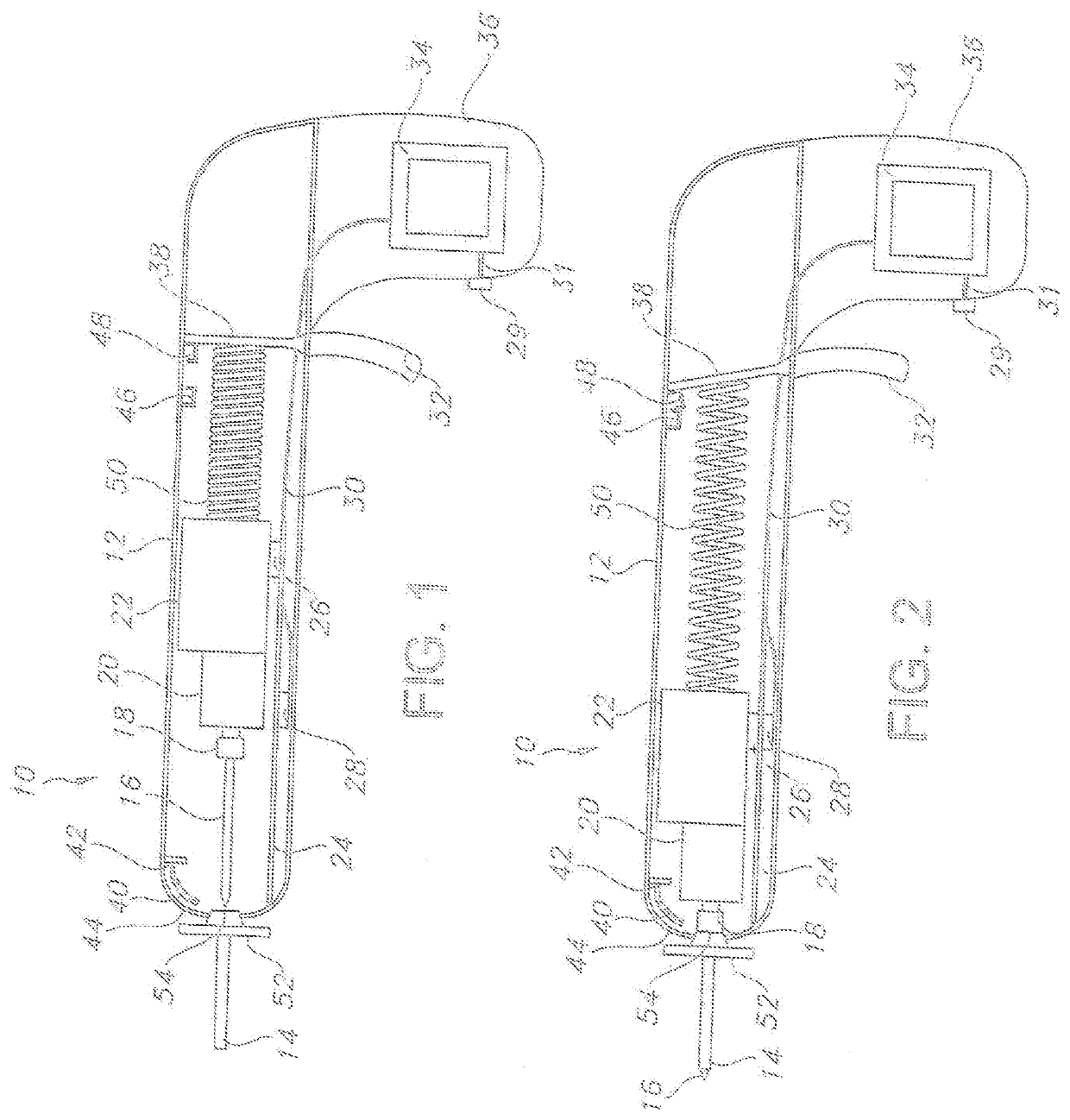
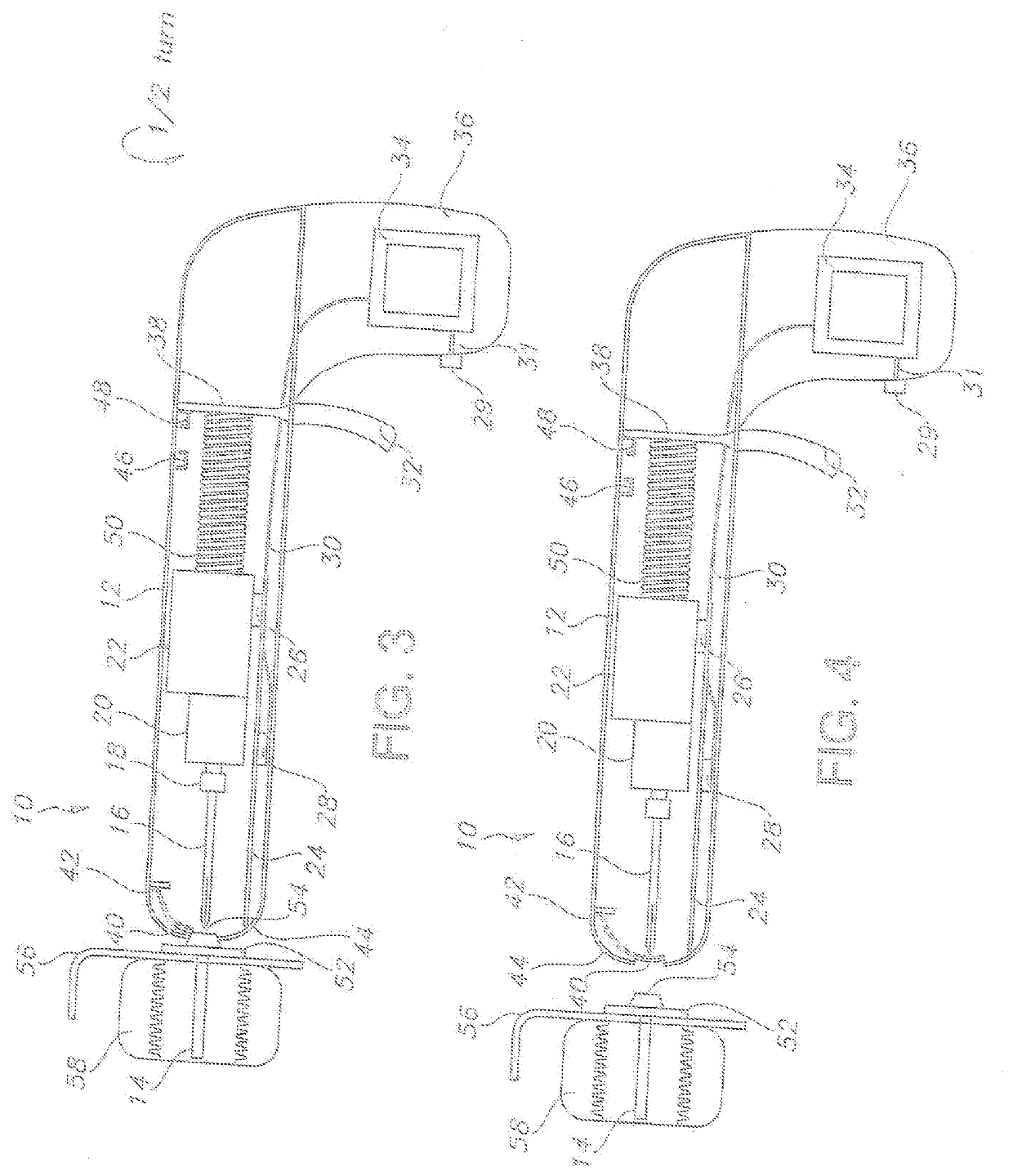
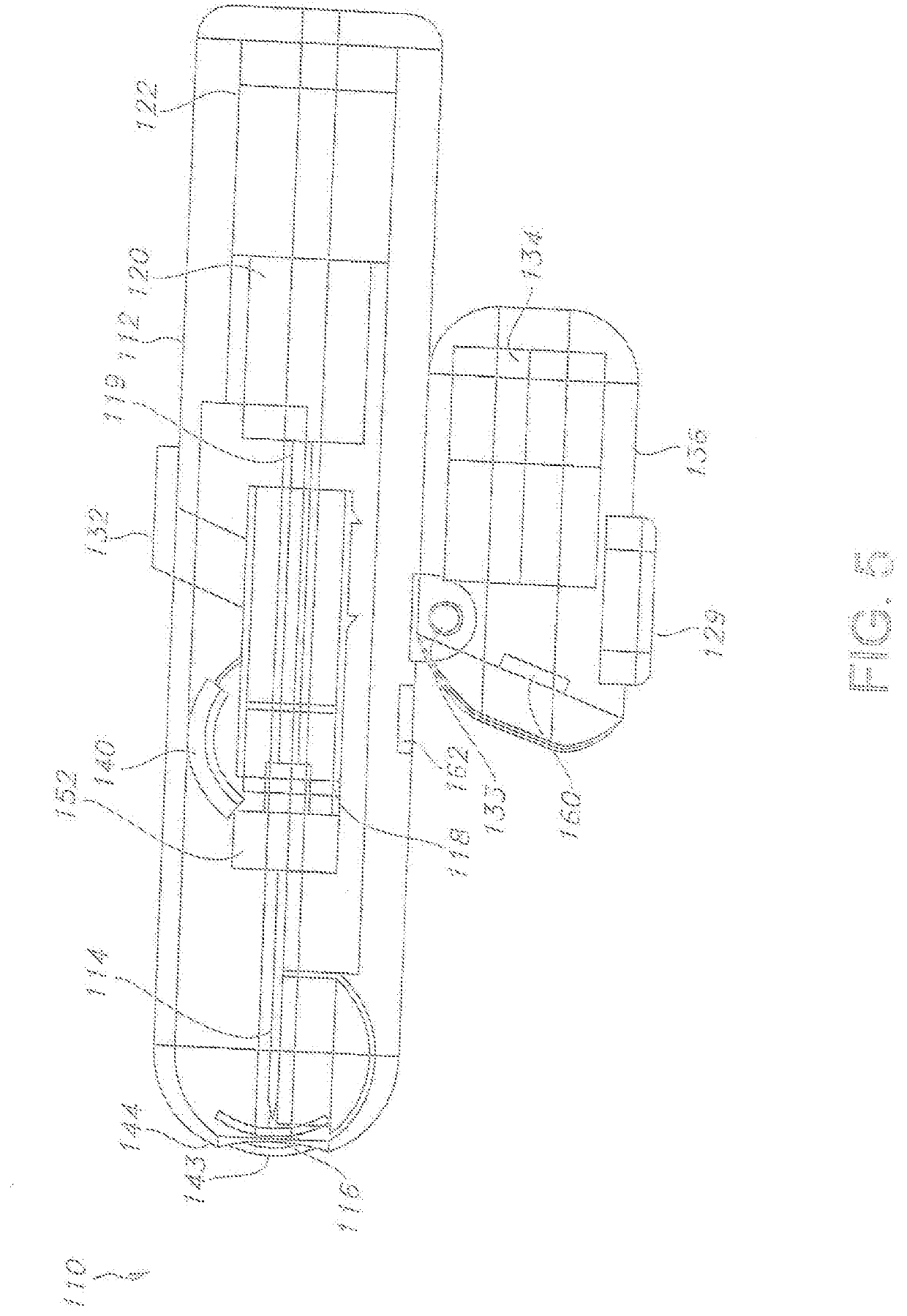
[0119]Reference now should be made to the drawings, presented as non-limiting possible embodiments in accordance with the descriptions provided above. The ordinarily skilled artisan would fully understand the breadth and scope intended herein in relation to the following potentially preferred types.

[0120]It will be understood that, although the terms first, second, third, etc. may be used herein to describe various elements, these elements should not be limited by these terms. These terms are only used to distinguish one element from another element. Thus, a first element discussed below could be termed a second element without departing from the teachings of the present disclosure.

[0121]The terminology used herein is for the purpose of describing particular embodiments only and is not intended to be limiting. As used herein, the singular forms “a”, “an”, and “the” are intended to include the plural forms as well, unless the context clearly indicates otherwise. It will be further un...


PUM


Abstract

A portable and passive safety intraosseous device to allow for direct introduction of medications, etc., within the intermedullary space of a subject patient's bone or, if needed, the removal of certain substances from such a subject patient's bone. Such a device permits direct drilling and placement of a cannula within the subject bone with access external to the subject patient's skin, permitting, as well, connection of a tube for such introduction/removal purposes. The ability to provide a passive safety unit allows for facilitated utilization in, for instance, emergency situations with the entire device provided for utilization thereof. The device includes a drilling component with a permanently attached stylet and a removable cannula, a power supply for a single drilling operation, a mechanism to draw the stylet back into the drill component after use and disengagement from the cannula, and an automatic closure that activates with the separation of the cannula.

Description

CROSS-REFERENCE TO RELATED APPLICATIONS[0001]This application is a continuation-in-part of pending U.S. patent application Ser. No. 16 / 125,767, filed on Sep. 10, 2018, which claims the benefit of expired U.S. Provisional Patent Application No. 62 / 556,397, filed on Sep. 9, 2017, and 62 / 566,498, filed on Oct. 1, 2017. The entirety of the parent application and both provisional applications are herein incorporated by reference.FIELD OF THE INVENTION[0002]The disclosure relates to a portable and passive safety intraosseous device to allow for direct introduction of medications, etc., within the intermedullary space of a subject patient's bone or, if needed, the removal of certain substances from such a subject patient's bone. Such a device permits direct drilling and placement of a cannula within the subject bone with access external to the subject patient's skin, permitting, as well, connection of a tube for such introduction / removal purposes. The ability to provide a passive safety un...

Claims


Application Information

Patent Timeline
Patent Type & Authority Applications(United States)
IPC IPC(8): A61B17/34A61B17/16
CPCA61B2017/00477A61B2017/00115A61B17/3496A61B17/3472A61B2017/00734A61B17/1628A61B17/3423A61B2017/0046A61B2017/291A61B2090/065A61B2090/0801A61B2090/309
Inventor COPPEDGE, BILLIEKARPOWICZ, EDWARDMARINELLI, STEVEN ANTHONY
Owner JUNE MEDICAL IP LLC
Why Eureka
Try Eureka