

Composition for diagnosis of degenerative neurological diseases
- Summary
- Abstract
- Description
- Claims
- Application Information
AI Technical Summary
Benefits of technology
Problems solved by technology
Method used
Image

Examples
Embodiment Construction
Technical Problem
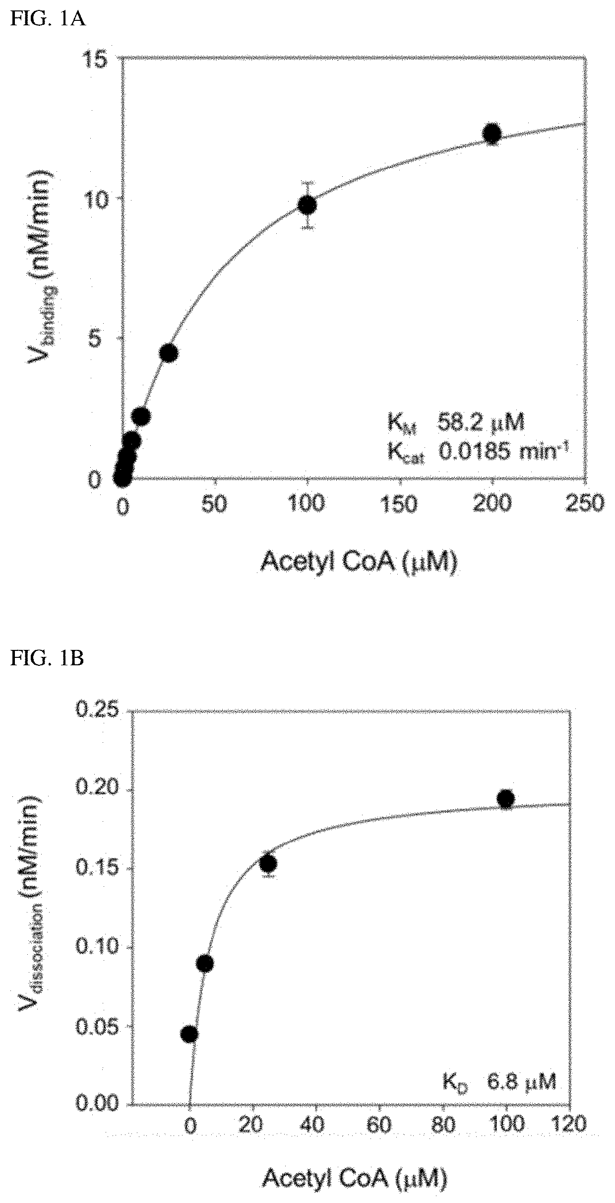
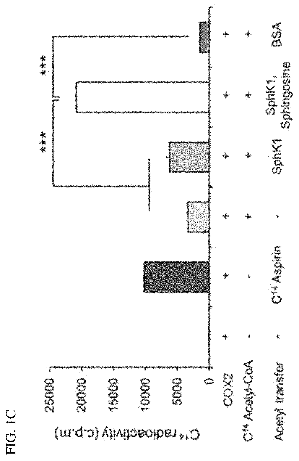
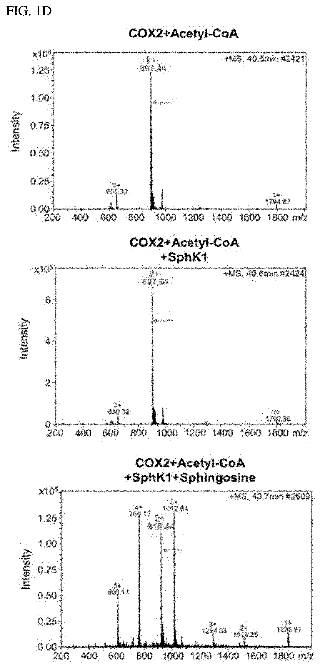
[0006]Accordingly, the present inventors made many efforts to find the role of SphK1 in degenerative neurological diseases, and as a result, found that SphK1 was exhibited by increasing the acetylation of cyclooxygenase 2 (COX2) to promote the secretion of neuroinflammatory resolution factor and resulting in inducing the conversion to M2-like phenotype of microglia. In addition, the present inventors confirmed that when the SphK1 was increased in the brain of AD, the acetylation of COX2 was increased, and as a result, Aβ phagocytosis through neuroglia cells was improved, thereby improving cognitive impairment. Furthermore, the present inventors found that the acetylation of COX2 was decreased in microglia and monocytes of an Alzheimer's animal model, which was exhibited at serine 565 of a COX2 protein, and completed the present invention.
[0007]Therefore, an object of the present invention is to provide a composition for diagnosis of degenerative neurological disease...
PUM
| Property | Measurement | Unit |
|---|---|---|
| Fraction | aaaaa | aaaaa |
Abstract
Description
Claims
Application Information