Systems and Methods for Joint Reconstruction and Segmentation of Organs From Magnetic Resonance Imaging Data

a magnetic resonance imaging and joint reconstruction technology, applied in the field of medical imaging and computer vision technology, can solve the problems of inability to fully sample mri scans, inability to use cardiac mri, and inability to accurately predict the effect of the patient's health, so as to achieve rapid static and dynamic reconstruction and segmentation.

Active Publication Date: 2021-04-22
RUTGERS THE STATE UNIV
View PDF0 Cites 6 Cited by
  • Summary
  • Abstract
  • Description
  • Claims
  • Application Information

AI Technical Summary

Benefits of technology

[0005]The present disclosure relates to systems and methods for joint reconstruction and segmentation of organs from magnetic resonance imaging (MRI) data. Sparse MRI data (k-space MRI data) is received at a computer system. The computer system jointly processes the MRI data using a plurality of reconstruction and segmentation processes. For example, the MRI data is processed using a joint reconstruction and segmentation process to identify an organ from the MRI data. Additionally, the MRI data is processed using a channel-wise attention network to perform static reconstruction of the organ from the MRI data. Further, the MRI data can be processed using a motion-guided network to perform dynamic reconstruction of the organ from the MRI data. Advantageously, the joint processing performed by the systems and methods of the present disclosure allow for rapid static and dynamic reconstruction and segmentation of organs from sparse MRI data, with particular advantage in clinical settings.

Problems solved by technology

Magnetic resonance imaging (“MRI”) is widely used due to its high resolution, high contrast, and no radiation, but a fully-sampled MRI scan requires a lot of time for a patient to undergo the scan.
One of the biggest hurdles to more extensive use of cardiac MRI is how long the exam takes (over an hour) and how complicated the procedures are (patients need to hold their breath during certain sequences).
The breath hold is necessary to cease any motion that might obscure the image, but not all patients are able to do this for the required length of time or repeatedly.
Because of the low contrast and large variation in intensity and shapes, myocardium segmentation has been a challenging task.

Method used


Image

  • Systems and Methods for Joint Reconstruction and Segmentation of Organs From Magnetic Resonance Imaging Data
  • Systems and Methods for Joint Reconstruction and Segmentation of Organs From Magnetic Resonance Imaging Data
  • Systems and Methods for Joint Reconstruction and Segmentation of Organs From Magnetic Resonance Imaging Data

Examples


Embodiment Construction

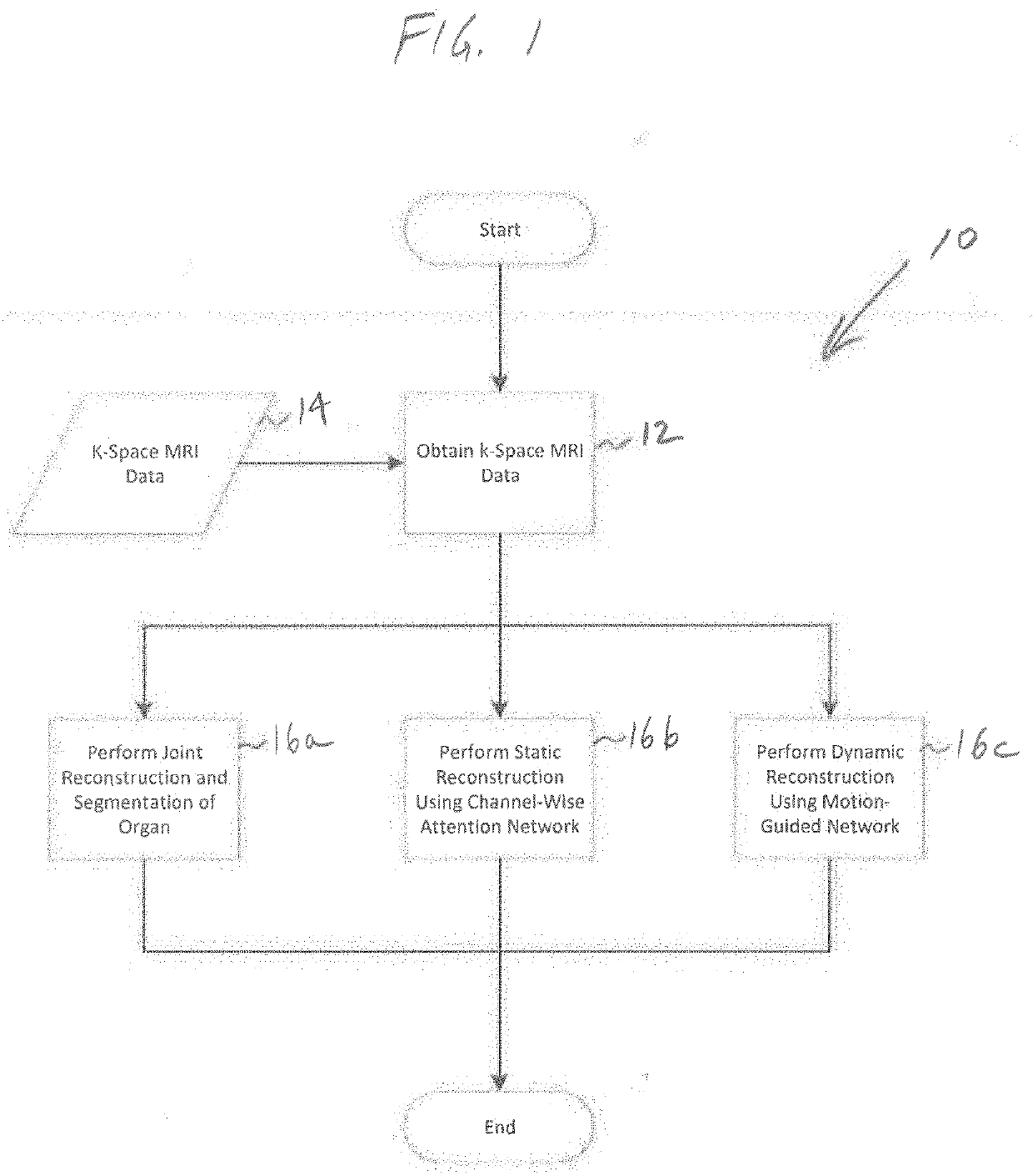
[0017]The present disclosure relates to systems and methods for joint reconstruction and segmentation of organs from MRI data, as discussed in detail below in connection with FIGS. 1-13.

[0018]FIG. 1 is a flowchart illustrating overall processing steps carried out by the systems and methods of the present disclosure, indicated generally at 10. In step 12, the system obtains k-space MRI data 14 from a suitable source, such as directly from an MRI scanner, from a computer system, and / or from a database of MRI data. The k-space MRI data 14 typically is undersampled data generated by the MRI machine when a scan is performed on a patient. In the past, the k-space MRI data 14 often did not have sufficient information to allow for adequate reconstruction and segmentation of organs in clinical applications, including both static and dynamic reconstruction and segmentation. Importantly, the three joint processes 16a-16c of the systems and methods of the present disclosure, discussed in more d...


PUM


Abstract

Systems and methods for joint reconstruction and segmentation of organs from magnetic resonance imaging (MRI) data are provided. Sparse MRI data is received at a computer system, which jointly processes the MRI data using a plurality of reconstruction and segmentation processes. The MRI data is processed using a joint reconstruction and segmentation process to identify an organ from the MRI data. Additionally, the MRI data is processed using a channel-wise attention network to perform static reconstruction of the organ from the MRI data. Further, the MRI data can is processed using a motion-guided network to perform dynamic reconstruction of the organ from the MRI data. The joint processing allows for rapid static and dynamic reconstruction and segmentation of organs from sparse MRI data, with particular advantage in clinical settings.

Description

RELATED APPLICATIONS[0001]The present application claims the benefit of U.S. Provisional Application Ser. No. 62 / 916,455 filed on Oct. 17, 2019, the entire disclosure of which is expressly incorporated herein by reference.BACKGROUNDTechnical Field[0002]The present disclosure relates generally to the field of medical imaging and computer vision technology. More specifically, the present invention relates to systems and methods for joint reconstruction and segmentation of organs from magnetic resonance imaging (“MRI”) data.Related Art[0003]Magnetic resonance imaging (“MRI”) is widely used due to its high resolution, high contrast, and no radiation, but a fully-sampled MRI scan requires a lot of time for a patient to undergo the scan. Such scans could greatly benefit from advances being made in machine learning techniques (artificial intelligence or “AI”) for MRI reconstruction of under-sampled MRI scan data that specifically benefit cardiac MRIs. In particular, there is a need to impr...

Claims


Application Information

Patent Timeline
Patent Type & Authority Applications(United States)
IPC IPC(8): G06T11/00G06T7/11G06T7/13G06T7/20
CPCG06T11/006G06T7/11G06T7/13G06T2211/412G06T2207/10088G06T2207/20084G06T2207/30048G06T7/20A61B5/055G01R33/5608G01R33/561G01R33/56325A61B5/0044A61B5/4887A61B5/7267A61B2576/023G16H30/40G16H50/20G06N3/084G06N3/044G06N3/045
Inventor HUANG, QIAOYINGMETAXAS, DIMITRIS
Owner RUTGERS THE STATE UNIV
Why Eureka
Try Eureka