
Multi-source medical imaging system

a multi-source, medical imaging technology, applied in the direction of instruments, static indicating devices, cathode-ray tube indicators, etc., can solve the problem of small areas of clinical or hospital setting cluttered

Inactive Publication Date: 2010-11-25
NDS SURGICAL IMAGING
View PDF19 Cites 47 Cited by
  • Summary
  • Abstract
  • Description
  • Claims
  • Application Information

AI Technical Summary

Benefits of technology

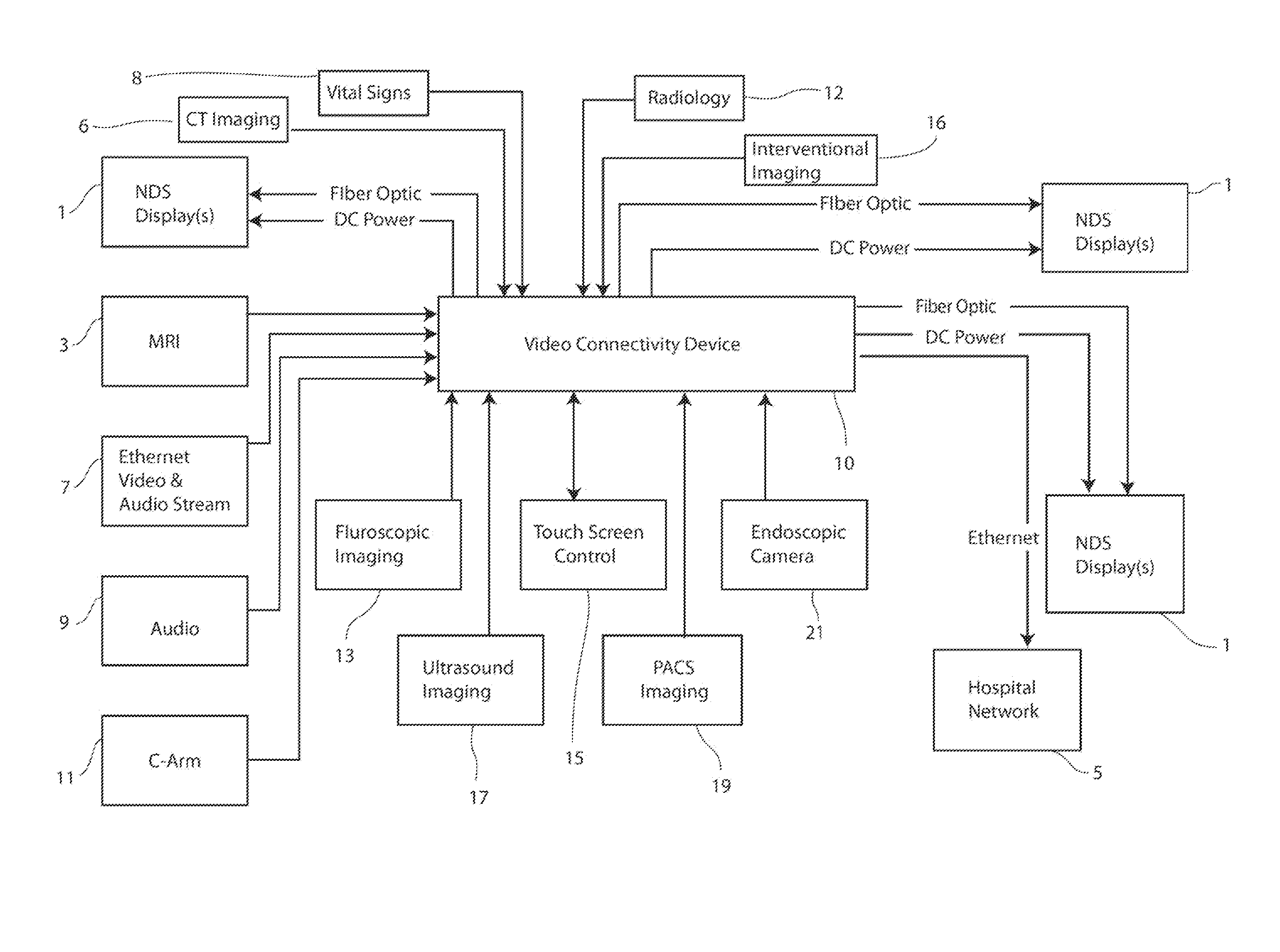
[0005]One aspect of the invention includes a multifunctional device that offers a complete video switching, video combining, image streaming and digital capture solution for surgical carts and / or integrated operating rooms (OR). This device offers connectivity between medical professionals by enabling real-time interactive consultation across rooms or continents. This device allows video and imaging signal control for surgical and interventional suites. Centralized DC power and fiber-based video delivery for displays makes OR installations using the device feasible in a hospital environment. The device brings video integration and distribution benefits to small and large surgical environments.
[0008]The video connectivity device can combine all input modalities and present them to a display that matches the resolution and attributes of the connectivity device output. The video connectivity device includes dual DVI and HD-SDI inputs as well as full multi-modality input combination flexibility. This embodiment allows any input to be viewed with any other input in a split-screen or Picture In Picture (PIP) configuration. In addition, the video connectivity device may be equipped with HD video streaming capabilities.

Problems solved by technology

This traditional signal routing mechanism causes clutter in small areas of a clinical or hospital setting.

Method used


Image

  • Multi-source medical imaging system
  • Multi-source medical imaging system
  • Multi-source medical imaging system

Examples


Embodiment Construction

[0029]One aspect of the invention relates to a video connectivity device for integrated operating rooms (ORs). The video connectivity device provides compliancy with ORs, hospitals and / or clinical settings, video control applications and control systems. The video connectivity device enables multi-modality imaging, integrating images from pathology, PACS imaging, fluoroscopy and ultrasound with surgical video. The video connectivity device further enables extended clinical reach and medical documentation. The video connectivity device is coupled to multiple displays (e.g., 23″ Radiance® surgical displays). Alternatively, multiple video connectivity devices may be coupled to one display.

[0030]An aspect of the invention is a video connectivity device that transmits output to multiple displays. The video connectivity device is a system intended for installation on surgical carts.

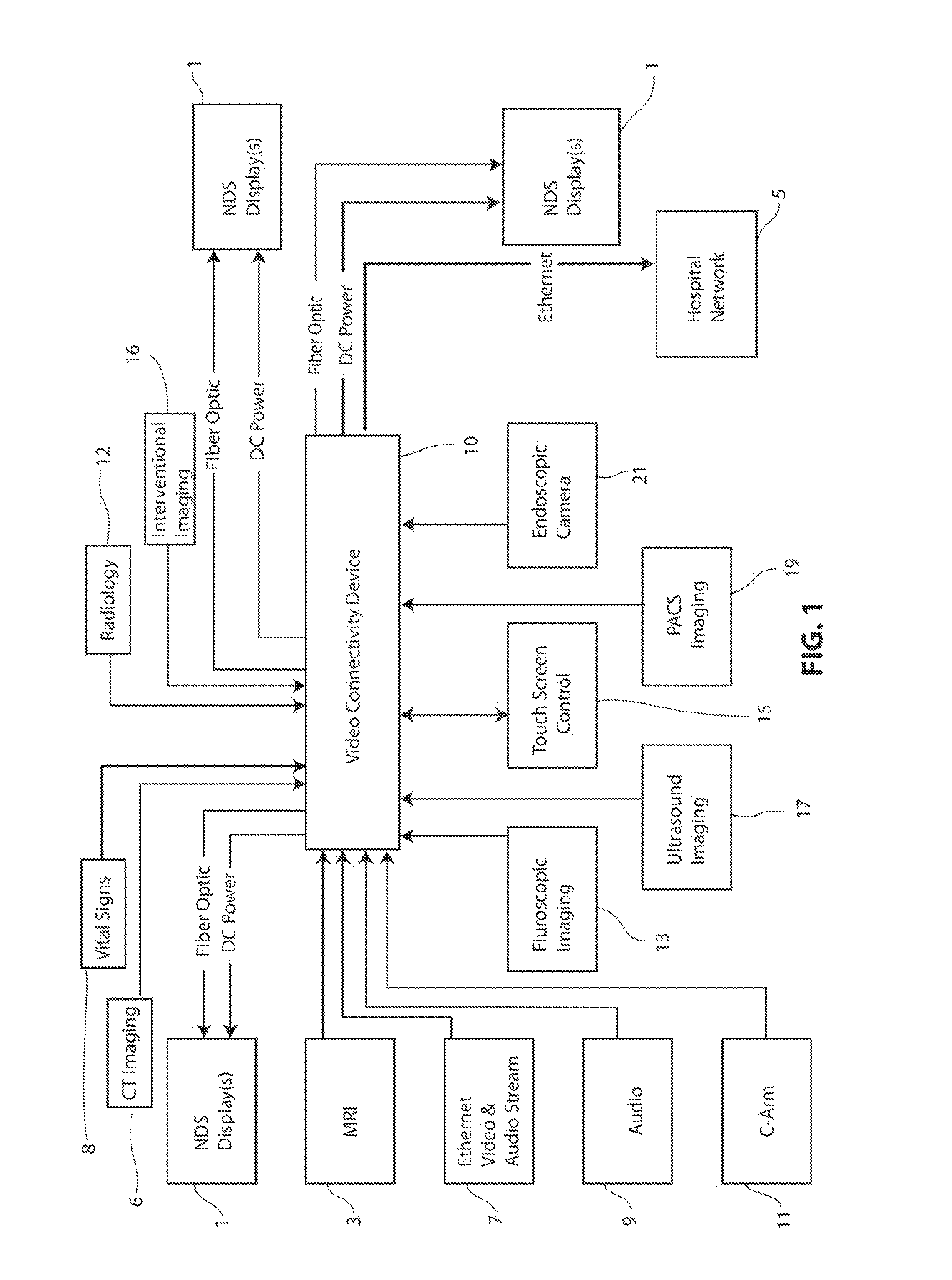
[0031]FIG. 1 illustrates one embodiment of a system for video distribution and telemedicine connectivity sup...


PUM


Abstract

This invention related to a clinical connectivity device coupled to a display. The clinical connectivity device comprises a user interface configured to select a display format; a controller configured to receive commands from the user interface and for controlling operation of the clinical connectivity device; and a selector coupled to the controller and an adjuster. The selector is configured to receive input signals of graphics, image, video, audio or data from devices in a clinical setting and is further configured to select a signal to be transmitted to an adjuster based on the input signal format. The adjuster is coupled to the controller and the display for matching the received signal format to the display format. The adjuster comprises a temporal combiner configured to combine synchronous and asynchronous received signals; a spatial combiner configured to place graphics, image, video or data; a scaler for geometric scaling and non-geometric scaling of graphics, image, video or data; and a rotation component configured to rotate graphics, image, video or data.

Description

CROSS REFERENCE TO RELATED APPLICATIONS[0001]This application claims priority to U.S. Provisional Patent Application No. 61 / 180,808 (filed May 22, 2009) entitled, “MULTI-SOURCE MEDICAL IMAGING SYSTEM” and U.S. Provisional Patent Application No. 61 / 251,201 (filed Oct. 13, 2009) entitled, “MULTI-SOURCE MEDICAL IMAGING SYSTEM.”INCORPORATION BY REFERENCE[0002]The subject matter of the present application is related to subject matter described in U.S. patent application Ser. No. 11 / 715,711 (filed Oct. 7, 2007) entitled, “WIDE VIEW DISPLAY SYSTEM FOR MEDICAL SURGICAL APPLICATIONS,” which are incorporated herein by reference. All publications and patent applications mentioned in this specification are herein incorporated by reference to the same extent as if each individual publication or patent application was specifically and individually indicated to be incorporated by reference.FIELD OF THE INVENTION[0003]The present invention relates generally to imaging devices used in medicine as we...

Claims


Application Information

Patent Timeline
Patent Type & Authority Applications(United States)
IPC IPC(8): G09G5/00
CPCG09G5/005G09G2380/08G09G2340/0435G09G2340/04
Inventor BAGHDADI, AMIRTRUPPA, JEROMECHANG, CHAD C.DUGGAN, GARY J.SAGHAFI, BABAKPILGUN, ALEXANDERHUANG, CHUNG-YINGFAN, YIYINGTHAI, MICHAELPOST, JONAHHANSEN, RONALD
Owner NDS SURGICAL IMAGING
Learn More