
Methods and systems for determining complementary ultrasound views

a complementary ultrasound and view technology, applied in the field of ultrasound, can solve the problems of long measurement time, manual measurement, and difficulty in manually selecting the appropriate additional view

Pending Publication Date: 2021-11-11
KONINKLJIJKE PHILIPS NV
View PDF0 Cites 0 Cited by
  • Summary
  • Abstract
  • Description
  • Claims
  • Application Information

AI Technical Summary

Benefits of technology

The patent text describes a way to simplify and speed up the process of fetal screening by using multiple views and options to analyze abnormal features. This helps to provide early detection and intervention for potential health risks to the fetus.

Problems solved by technology

Manually selecting the appropriate additional views to observe the anatomy and manually performing the measurements can be long and difficult, especially if several anatomical regions need to be examined.

Method used


Image

  • Methods and systems for determining complementary ultrasound views
  • Methods and systems for determining complementary ultrasound views
  • Methods and systems for determining complementary ultrasound views

Examples


Embodiment Construction

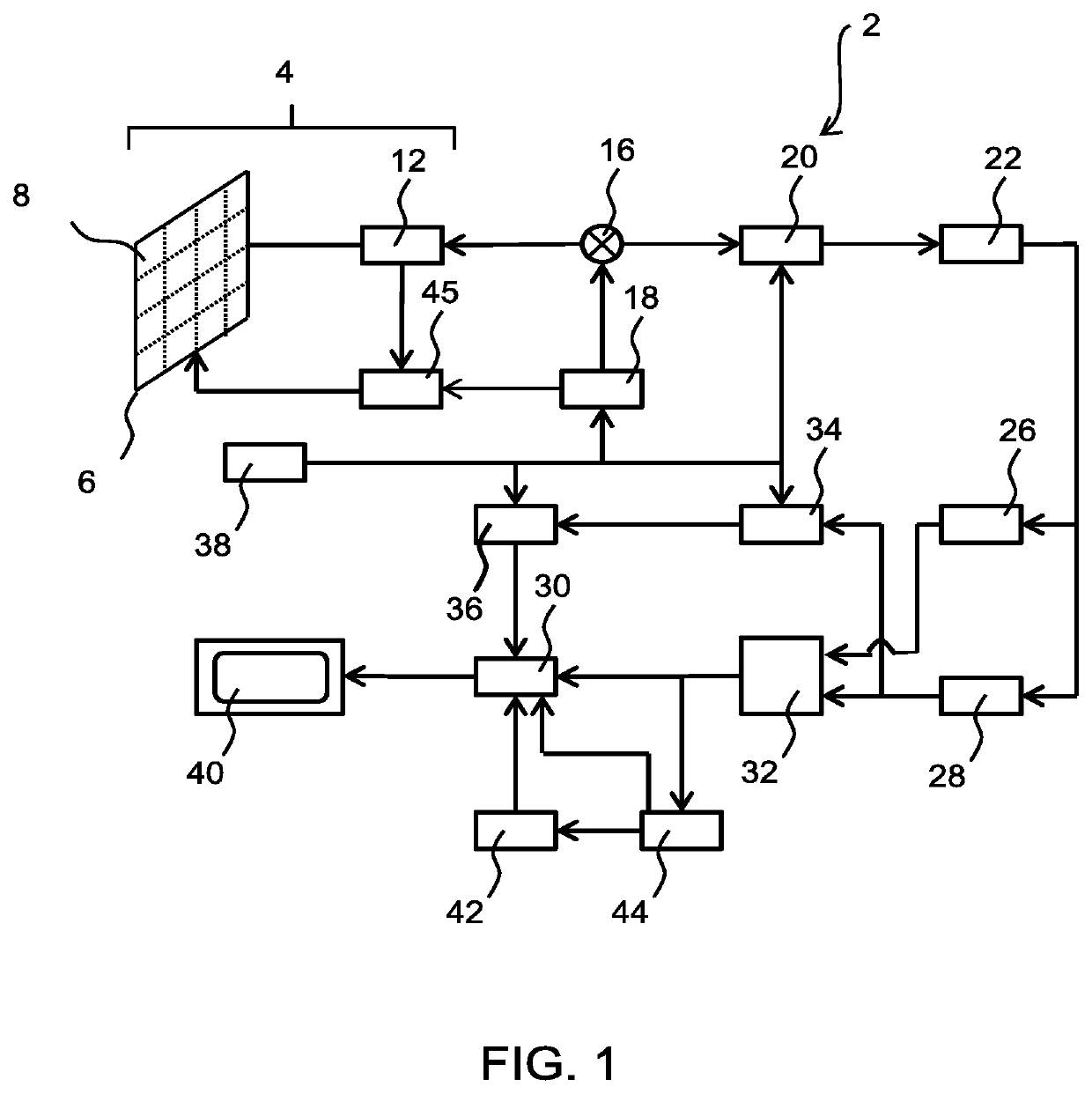
[0072]The invention will be described with reference to the Figures.

[0073]It should be understood that the detailed description and specific examples, while indicating exemplary embodiments of the apparatus, systems and methods, are intended for purposes of illustration only and are not intended to limit the scope of the invention. These and other features, aspects, and advantages of the apparatus, systems and methods of the present invention will become better understood from the following description, appended claims, and accompanying drawings. It should be understood that the Figures are merely schematic and are not drawn to scale. It should also be understood that the same reference numerals are used throughout the Figures to indicate the same or similar parts.

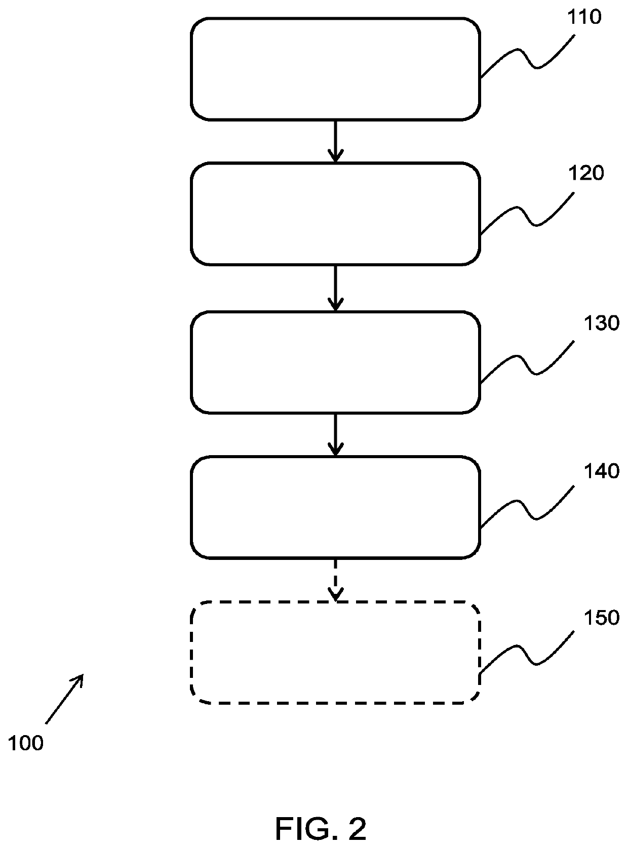
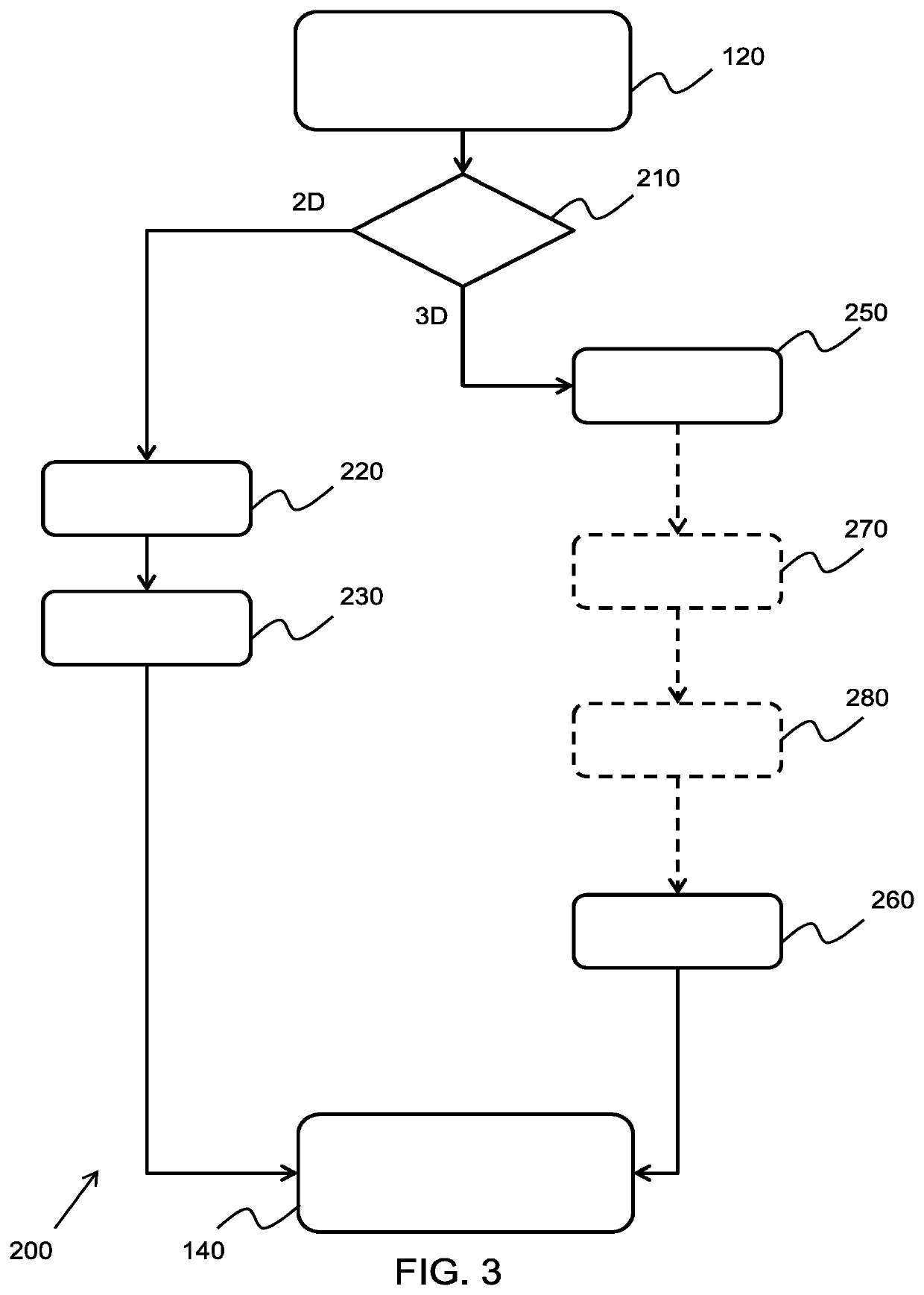
[0074]The invention provides an ultrasound imaging method for determining complementary views of interest based on an anomalous feature identified in a region of interest of an ultrasound image. The method includes obtaini...


PUM


Abstract

The invention provides an ultrasound imaging method for determining complementary views of interest based on an anomalous feature identified in a region of interest of an ultrasound image. The method includes obtaining an ultrasound image of a region of interest of a subject and identifying an anomalous feature within said region. The identified anomalous feature may then be used to determine one or more available complementary ultrasound images of interest of the subject. The one or more available complementary ultrasound images may then be displayed to a user and the complementary ultrasound views to be reviewed may then be selected by the user from the displayed available complementary ultrasound images.

Description

FIELD OF THE INVENTION[0001]This invention relates to the field of ultrasound, and in particular to the field of ultrasound imaging.BACKGROUND OF THE INVENTION[0002]Ultrasound imaging is used in a wide array of medical imaging applications. When performing ultrasound imaging, it is possible that anomalies may be detected, which may be indicative of an underlying medical condition.[0003]A common application of ultrasound imaging is a fetal screening exam, where the health of a fetus is assessed during a given stage in the pregnancy. During the screening exam, the clinician performs measurements of various specific anatomical structures and compares the values to common measurement tables. When a measurement is not in the normal range, additional examinations and additional measurements are required to confirm that the anomaly is a cause for concern. The additional examinations and measurements typically vary depending on the suspected pathology.[0004]Manually selecting the appropriat...

Claims


Application Information

Patent Timeline
Patent Type & Authority Applications(United States)
IPC IPC(8): A61B8/08A61B8/00
CPCA61B8/085A61B8/463A61B8/5223A61B8/0866A61B8/466A61B8/145A61B8/483A61B8/523
Inventor CIOFOLO-VEIT, CYBÈLELEFEVRE, THIERRYRAYNAUD, CAROLINE DENISE FRANCOISEROUET, LAURENCE
Owner KONINKLJIJKE PHILIPS NV
Learn More