Continuous and dynamic ejection fraction determination

a technology of ejection fraction and determination method, which is applied in the field of systolic function of the heart, can solve the problems of delayed treatment of patients, significant worsening of disease conditions, and inaccurate assessment of current ejection fraction measurement methods

Pending Publication Date: 2022-04-07
CAPTION HEALTH INC
View PDF6 Cites 3 Cited by
  • Summary
  • Abstract
  • Description
  • Claims
  • Application Information

AI Technical Summary

Benefits of technology

[0006]Embodiments of the present invention address deficiencies of the art in respect to the determination of ejection fraction and provide a novel and non-obvious method, system and computer program product for the continuous and dynamic automated computation of ejection fraction. In an embodiment of the invention, a method for continuously and dynamically computing ejection fraction includes first training a neural network with different sets of cardiac imaging data acquired of a ventricle for different hearts and a known ejection fraction for each of the sets and then loading the trained neural network into memory of a computer. Afterwards, contemporaneous sets of imaging data of a ventricle of a heart may be continuously acquired according to a specified view. For each corresponding one of the sets of imaging data, an image quality value may then be computed for the corresponding one of the sets, and the corresponding one of the sets of imaging data may be provided to the neural network. The neural network, in response, provides, as output, an ejection fraction determination output without tracing a ventricle boundary of the heart. Thereafter, both the computed image quality value and the ejection fraction determination output may be displayed in a display of the computer.

Problems solved by technology

Current ejection fraction measurement methods tend to inaccurately assess disease conditions of the heart.
This error can lead to the delayed treatment of patients, and the significant worsening of disease conditions during the delay.
But, in doing so, the ventricle border needs to be manually traced by a human reader, or automatically traced using endocardial border detection methods, both of which methods are subject to error.
Moreover, this methodology relies on finding the exact end systolic and end diastolic image frames, often nontrivial step that can introduce errors if not done accurately.
Three dimensional echocardiograms may rely less on assumptions about the shape of the ventricle for calculating ejection fraction than two-dimensional mode, but it still requires the manual or automated and error-prone detection of endocardial borders.
As a result, current methods in ejection fraction measurement perform measurements in a way that is neither optimized nor reproducible despite the advances in modern diagnostic technologies.

Method used


Image

  • Continuous and dynamic ejection fraction determination
  • Continuous and dynamic ejection fraction determination

Examples


Embodiment Construction

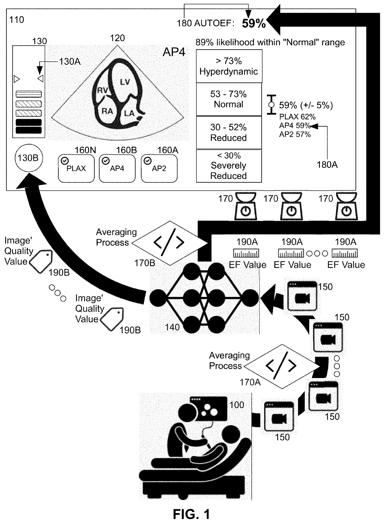
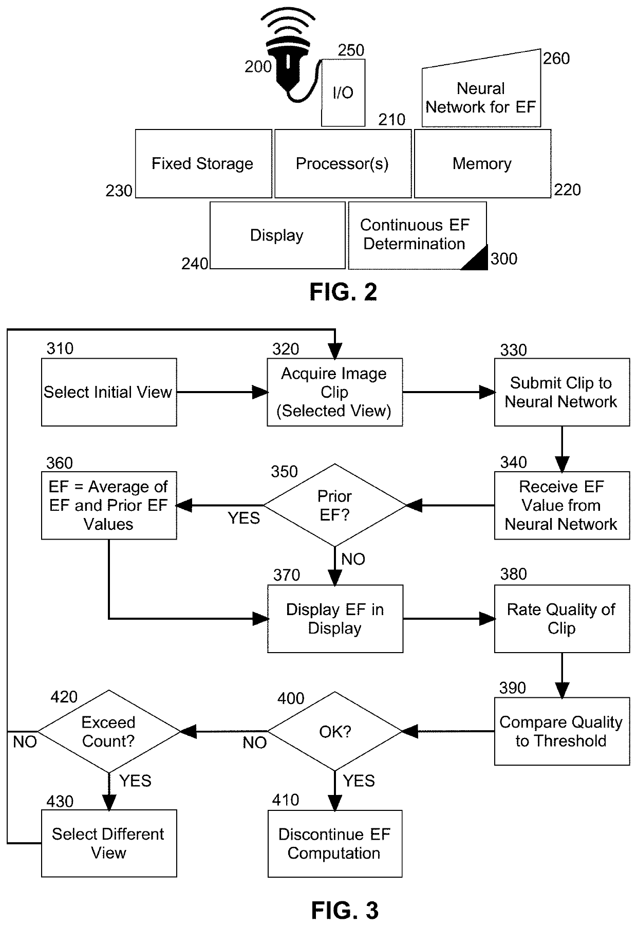
[0016]Embodiments of the invention provide for the continuous and dynamic computation of ejection fraction in a data processing system. In accordance with an embodiment of the invention, utilizing one or more views of a heart, different sets of contemporaneous imagery of the heart or a portion thereof are acquired ultrasonically and submitted to a neural network trained to correlate different sets of imagery of the heart with different ejection fraction values without requiring a tracing of the boundary of any portion of the heart. The neural network then produces a corresponding ejection fraction value for each submitted set of imagery which may then be displayed in a display of a computer. In this way, rather than receiving merely a finalized value for ejection fraction from a single set of acquired imagery of the heart, a continuously display of ejection fraction determinations may be provided in real-time as the heart is subjected to ultrasound imaging such that an operator may ...


PUM


Abstract

The continuous and dynamic computation of ejection fraction (EF) includes training a neural network with different sets of cardiac imaging data acquired of a ventricle for different hearts and a known EF for each of the sets and then loading the trained neural network into memory of a computer. Afterwards, contemporaneous sets of imaging data of a ventricle of a heart are continuously acquired according to a specified view. For each corresponding set of imaging data, an image quality value may then be computed, and the corresponding set of imaging data may be provided to the neural network. The neural network, in response, provides, as output, an EF determination output without tracing a ventricle boundary of the heart. Thereafter, both the computed image quality value and the EF determination output may be displayed in a display of the computer.

Description

BACKGROUND OF THE INVENTIONField of the Invention[0001]The present invention relates to the systolic function of the heart and more particularly ejection fraction measurement.Description of the Related Art[0002]The ejection fraction is a measurement of the systolic function of the heart that refers to the percentage of blood leaving the heart at each contraction. Specifically, during each pumping cycle of the heart, the heart both contracts and also relaxes. When the heart contracts, the heart ejects blood from its two pumping chambers, known as the left ventricle and the right ventricle. Conversely, when the heart relaxes, both ventricles refill with blood. Of note, no matter how forceful the contraction of the heart, the heart does not pump all of the blood out of each ventricle. Instead, some blood remains. Hence, the term “ejection fraction” refers to the percentage of blood that is pumped out of a filled ventricle with each heartbeat.[0003]Of the two ventricles, the left ventri...

Claims


Application Information

Patent Timeline
Patent Type & Authority Applications(United States)
IPC IPC(8): A61B8/06A61B8/08A61B8/00G06T5/50
CPCA61B8/065A61B8/5207A61B8/461G06T5/50G06T2207/10132G06T2207/20084G06T2207/30048G06T2207/20216G06T2207/30168G06T2207/20081A61B8/0883A61B8/5223A61B8/463
Inventor CADIEU, CHARLESHONG, HAKOEPSELL, KILIANCHAUDHRY, ALIPOILVERT, NICOLASCANNON, MICHAEL G.PARAJULI, NRIPESH
Owner CAPTION HEALTH INC
Why Eureka
Try Eureka