Device for heart measurement

a heart measurement and heart valve technology, applied in the field of devices for heart measurement, can solve the problems of fatigue of afflicted patients, inability to perform even mild exertion tasks, pain and discomfort of patients, and the heart becomes so large that the heart cannot adequately supply blood

Inactive Publication Date: 2003-06-10
MARDIL
View PDF60 Cites 90 Cited by
  • Summary
  • Abstract
  • Description
  • Claims
  • Application Information

AI Technical Summary

Problems solved by technology

In time, the heart becomes so enlarged the heart cannot adequately supply blood.
An afflicted patient is fatigued, unable to perform even mildly exerting tasks and experiences pain and discomfort.
Further, as the heart enlarges, the internal heart valves may not adequately close.
This impairs the function of the valves and further reduces the heart's ability to supply blood.
This is a radical, new, experimental procedure that is subject to substantial controversy.
Furthermore, the procedure is highly invasive, risky and expensive and commonly includes other expensive procedures (such as a concurrent heart valve replacement).
Even though cardiomyoplasty has demonstrated symptomatic improvement, studies suggest the procedure only minimally improves cardiac performance.
The procedure is highly invasive requiring harvesting a patient's muscle and an open chest approach (i.e., sternotomy) to access the heart.
Furthermore, the procedure is expensive, especially when a paced muscle is utilized, because a costly pacemaker is required.
The cardiomyoplasty procedure is complicated.
For example, it is difficult to adequately wrap the muscle around the heart and attain a satisfactory fit.
Also, if adequate blood flow is not maintained to the wrapped muscle, the muscle may necrose.
The muscle may stretch after wrapping, reducing its constraining benefits, and the muscle is generally not susceptible to post-operative adjustment.
Finally, the muscle may fibrose and adhere to the heart causing undesirable constraint on the contraction of the heart during systole.
While cardiomyoplasty has resulted in symptomatic improvement, the nature of the improvement is not understood.
Such surgeries and devices are expensive.
The devices are at risk of mechanical failure and frequently require external power supplies.

Method used


Image

  • Device for heart measurement
  • Device for heart measurement
  • Device for heart measurement

Examples


second embodiment

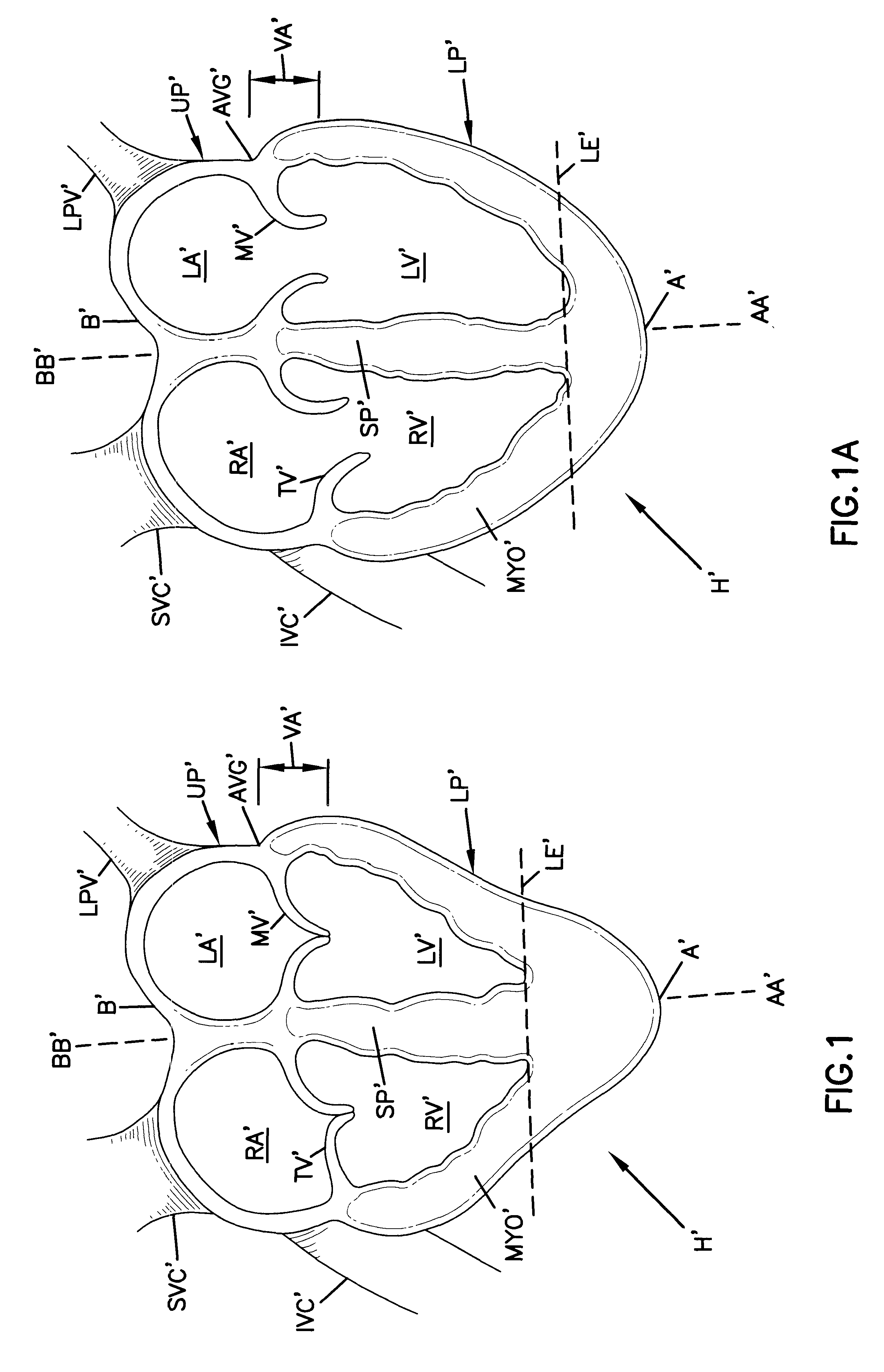
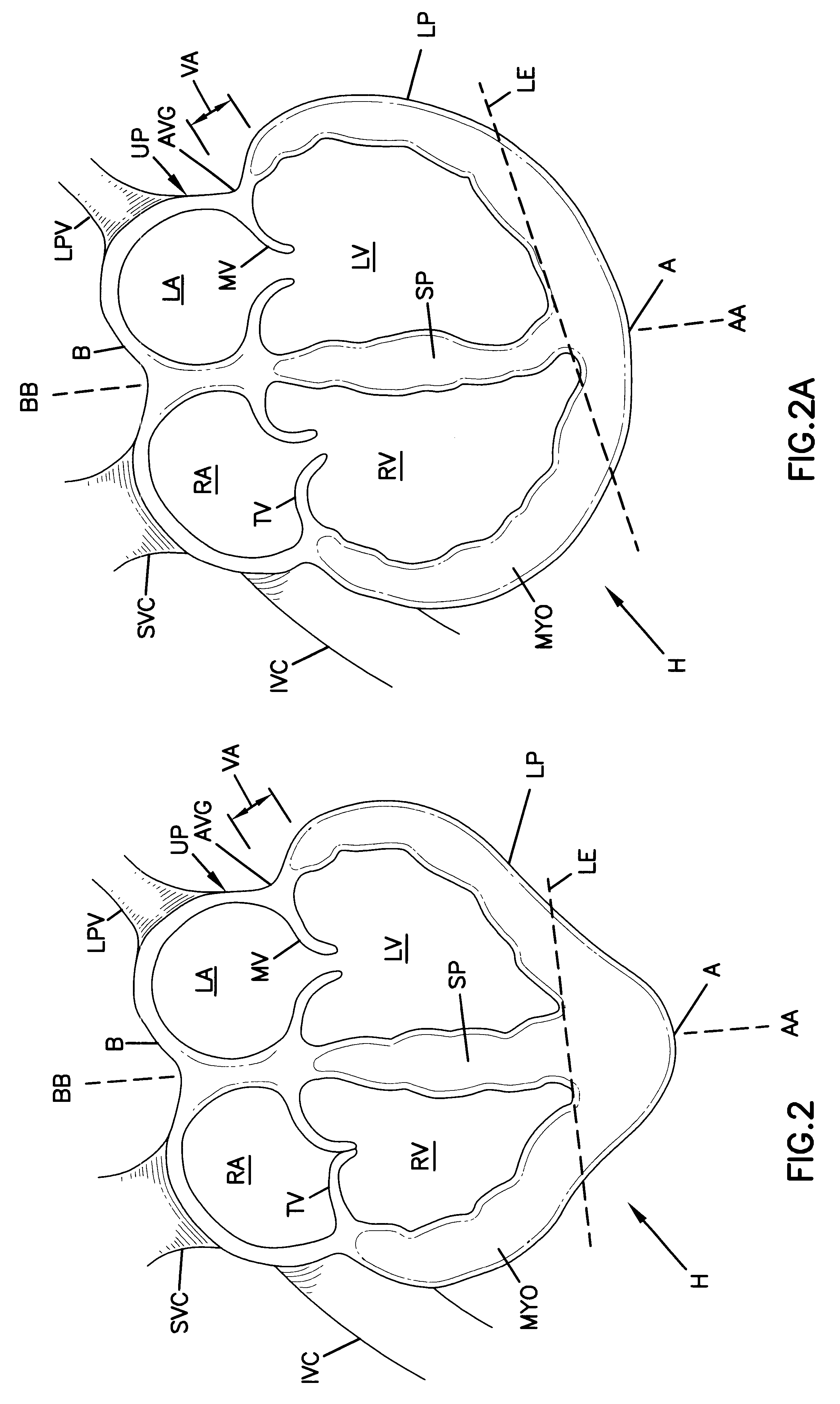
With reference now to FIGS. 3, 3A, 4 and 4A, the cardiac constraint device is shown as a jacket 10, 10' of flexible, biologically compatible material. The jacket 10, 10' is an enclosed knit material having upper and lower ends 12, 12', 14, 14'. The jacket 10, 10' defines an internal volume 16, 16' which is completely enclosed but for the open ends 12, 12' and 14'. In the embodiment of FIG. 3, lower end 14 is closed. In the embodiment of FIG. 4, lower end 14' is open. In both embodiments, upper ends 12, 12' are open. Throughout this description, the embodiment of FIG. 3 will be discussed. Elements in common between the embodiments of FIGS. 3 and 4 are numbered identically with the addition of an apostrophe to distinguish the second embodiment and such elements need not be separately discussed.

The jacket 10 is dimensioned with respect to a heart H to be treated. Specifically, the jacket 10 is sized for the heart H to be constrained within the volume 16. The jacket 10 can be slipped ar...

first embodiment

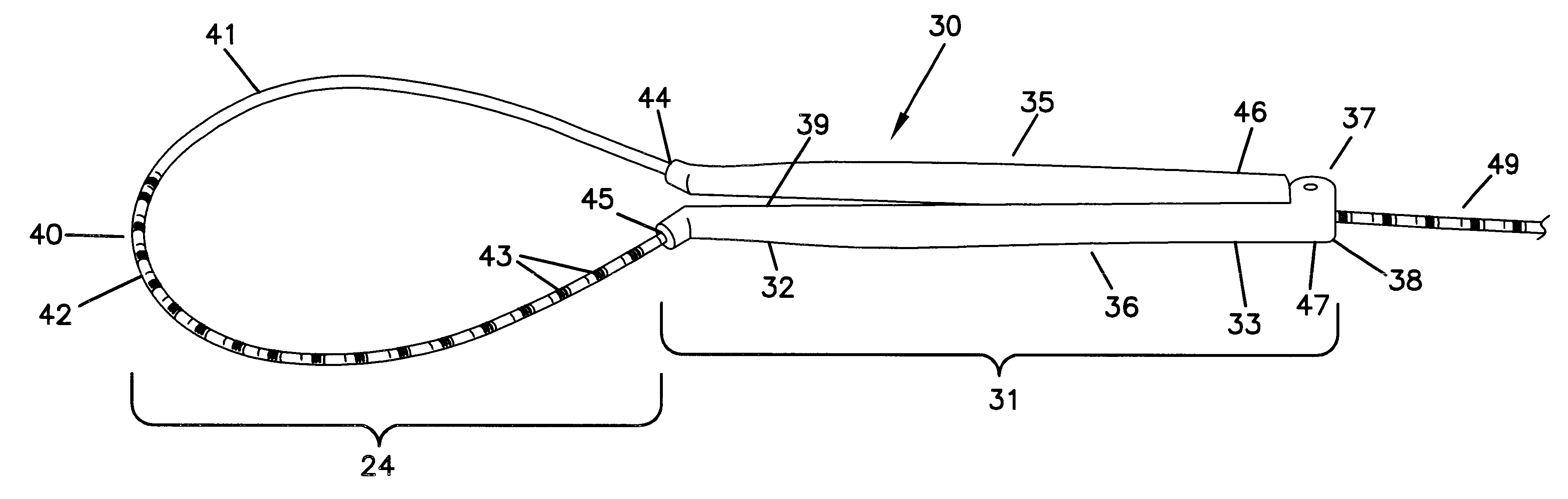
According to this embodiment, the diameter of the lumen 120 and 121 are constant along the length of the first and second handle members 110 and 111. In contrast to the first embodiment, the first handle member 110 does not have a lumen 120 configured to anchor the flexible member 101. Instead, the flexible member 101 is configured to be anchored within the lumen 120 of the first handle member 110. If desired, the handle members 110 and 111 can be identical and interchangeable.

As with the first embodiment, the first and second handle members 110 and 111, respectively, can be made of any material that is sterilizable. Preferably, the first and second handle members 110 and 111 are made of materials typically used in the manufacturing of medical devices. Examples of materials that could be used to make the first and second handle members 110 and 111 include plastics, such as polyethlyne, annealed stainless steel, brass or aluminum. If desired, the flexible member 101 and the first and...


PUM


Abstract

A surgical tool for measuring a heart that includes a first and second handle member having proximal and distal ends and internal passageways that run lengthwise from the proximal end to the distal end, a flexible member that is cylindrically shaped with a marked proximal end that is larger in diameter than the unmarked distal end, a hinged region with a connection receiving portion on the first handle member and a connection portion on the second handle member, wherein the distal end of the flexible member passes through the internal passageway of the first handle member from the proximal end of the first handle member to the distal end of the first handle member, extends out, then enters into the distal end of the second handle member, passes through the internal passageway of the second handle member from the proximal end to the distal end of the second handle member.

Description

I. BACKGROUND OF THE INVENTION1. Field of the InventionThe present invention pertains to a method and apparatus for treating congestive heart disease and related valvular dysfunction. More particularly, the present invention is directed to a measurement device for determining an appropriate size for a cardiac reinforcement device for a particular heart.2. Description of the Prior ArtCongestive heart disease is a progressive and debilitating illness. The disease is characterized by a progressive enlargement of the heart.As the heart enlarges, it is forced to perform an increasing amount of work in order to pump blood in each heart beat. In time, the heart becomes so enlarged the heart cannot adequately supply blood. An afflicted patient is fatigued, unable to perform even mildly exerting tasks and experiences pain and discomfort. Further, as the heart enlarges, the internal heart valves may not adequately close. This impairs the function of the valves and further reduces the heart's ...

Claims


Application Information

Patent Timeline
Patent Type & Authority Patents(United States)
IPC IPC(8): A61B5/107
CPCA61B5/107A61F2/2481A61B5/1076A61B5/6869A61F2/2496
Inventor VANDEN HOEK, JOHNRIVERS, JODY
Owner MARDIL
Why Eureka
Try Eureka