Electrochemical sensor using intercalative, redox-active moieties

a technology of redox-active moieties and electrochemical sensors, applied in the field of electrochemical sensors using intercalative, redox-active moieties, can solve the problems of mismatch at the mutated site, severe consequences, and limited usefulness of a particular system for detecting materials of interest,

Inactive Publication Date: 2003-11-18
CALIFORNIA INST OF TECH
View PDF10 Cites 13 Cited by
  • Summary
  • Abstract
  • Description
  • Claims
  • Application Information

AI Technical Summary

Benefits of technology

The present invention provides a highly sensitive and accurate method for the detection of genetic point mutations and common DNA lesions in nucleic acid sequences and its application as a biosensor by DNA-mediated charge transport. The invention also provides an electrochemical assay of protein binding to DNA-modified electrodes based upon the detection of associated perturbations in DNA base stacking using DNA charge transport. In a particular embodiment, the invention relates to electrodes that are prepared by modifying their surfaces with oligonucleotide duplexes combined with an intercalative, redox-active species and their use as sensors based on an electrochemical process in which electrons are transferred between the electrode and the redox-active species. In another embodiment, the invention also relates to detection of base-pair mismatches, base flipping, or other base-stacking perturbations due to protein-nucleic acid interactions. Additionally, the protein-DNA interaction allows a method of electrical monitoring of DNA enzymatic reactions in a duplex nucleic acid sequence.

Problems solved by technology

It is now well known that mutations in DNA can lead to severe consequences in metabolic functions (e.g., regulation of gene expression, modulation of protein production) which ultimately are expressed in a variety of diseases.
However, the specific labeling method chosen depends on a multitude of factors, such as ease of attachment of the label, its sensitivity and stability over time, rapid and easy detection and quantification, as well as cost and safety issues.
Thus, despite the abundance of labeling techniques, the usefulness, versatility and diagnostic value of a particular system for detecting a material of interest is often limited.
Introduction of a mutation into double-stranded sequences creates a mismatch at the mutated site that destabilizes the DNA duplex.
The presence of a mismatch results in incomplete hybridization and thus cleavage by RNase A at the mutation site.
While these techniques are less sensitive than sequencing, they are simpler to perform on a large number of tumor samples.
Each of these known prior art methods for detecting base pair mismatches has limitations that affect adequate sensitivity, specificity and ease of automation of the assay.
In particular, these methods are unable to detect mismatches independent of sequence composition and require carefully controlled conditions, and most methods detect multiple mismatches only.
Additional shortcomings that limit these methods include high background signal, poor enzyme specificity, and / or contamination.
Perturbations caused by mismatches or bulges greatly diminished the yields of DNA-mediated charge transport.
In the DNA double helix however, .pi.-stacking can contribute to electronic coupling such that small energetic differences could lead to large differences in coupling efficiency.
In general, owing to their relatively high thermodynamic stability and their minor modification to the DNA duplex, these lesions have been very difficult to detect, with postlabeling procedures the preferred methods of analysis.
These lesions do, however, appear to perturb electronic coupling within the DNA duplex.
This approach may be particularly useful to monitor assays based on redox-active proteins which bind to the oligonucleotide-modified surface, but are not easily oxidized or reduced because the redox-active center is not intercalating.
Furthermore, due to its catalytic nature, the measured charge increases with increased sampling times, and in principle is limited only by the concentration of ferricyanide in solution.
Electrodes with attenuated signals indicate the presence of the protein in question which is binding to its recognition site, thus causing a perturbation in base-stacking.
Third, the stability of the bond between the support and the immobilized molecule must be such to avoid leaching which would lead to reduced accuracy and sensitivity.

Method used


Image

  • Electrochemical sensor using intercalative, redox-active moieties
  • Electrochemical sensor using intercalative, redox-active moieties
  • Electrochemical sensor using intercalative, redox-active moieties

Examples


example 1

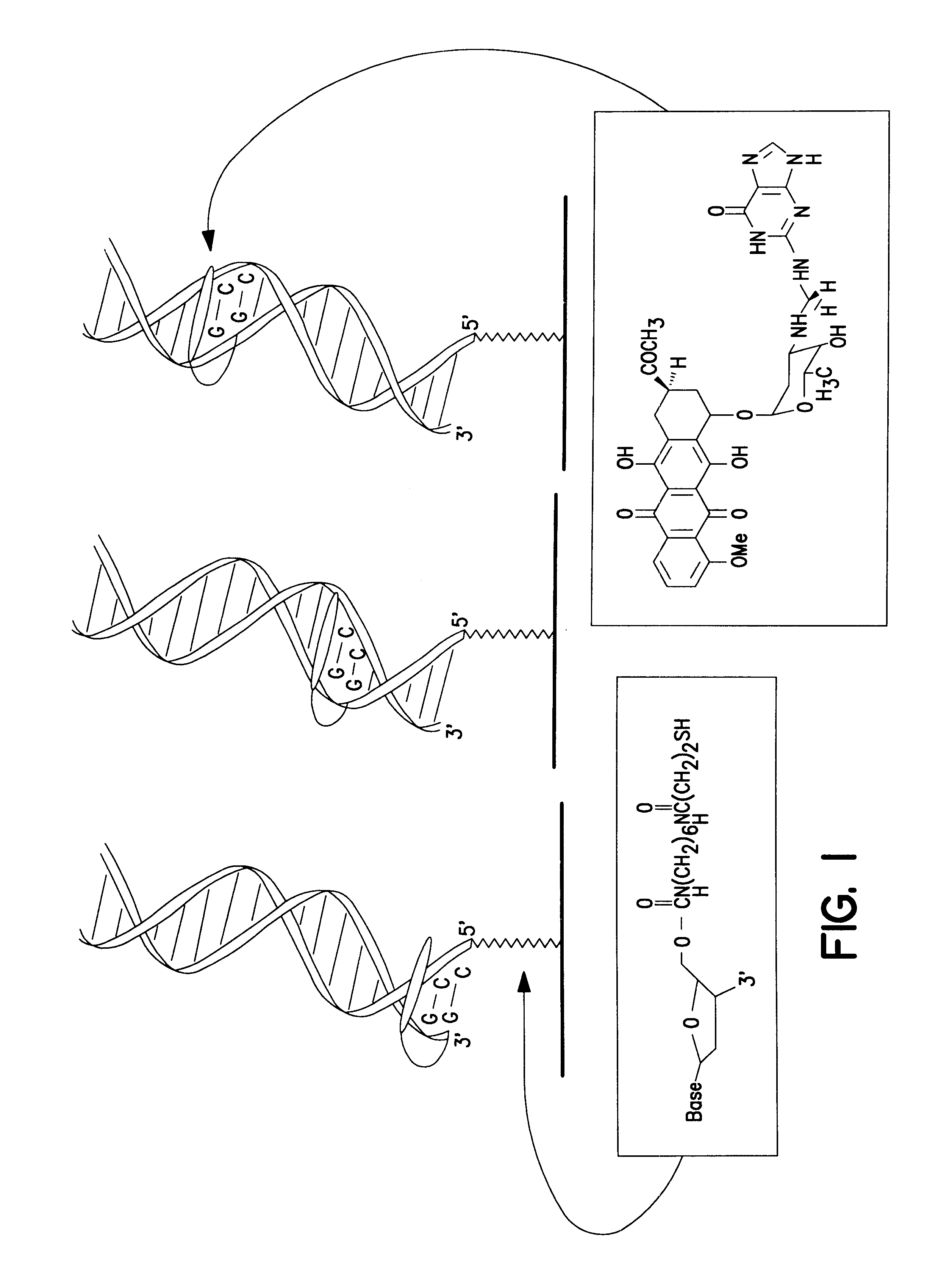
Site-Specific Incorporation of a Redox-Active Intercalator Into a DNA Duplex.

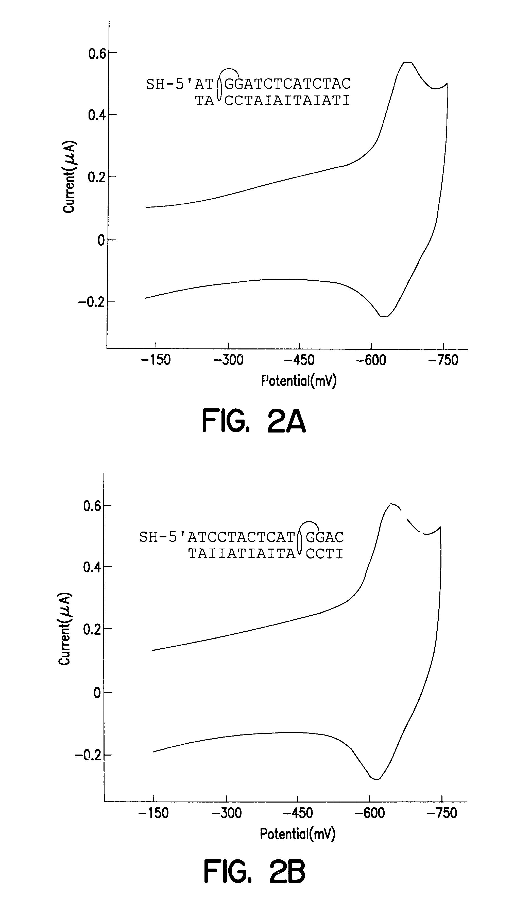
The redox-active intercalator daunomycin (DM) (Arcamone, 1981) was incorporated into the DNA duplex to investigate charge transduction through these duplexes (FIG. 1). DM undergoes a reversible reduction (Molinier-Jumel, 1978; Berg, 1981) within the potential window of the monolayers (Kelley, 1997a), and covalent adducts of intercalated DM crosslinked to the 2-amino group of guanine (Leng, 1996) have been crystallographically characterized within duplex DNA (Wang, 1991). Thus, a series of oligonucleotides primarily containing A-T or inosine (I)-C pairs were constructed with discrete guanine binding sites to which DM was crosslinked. Preferably, thiol-terminated duplexes (0.1 mM) containing an adjacent pair of guanines were hybridized, incubated with 0.2% formaldehyde and 0.2 mM DM in 5 mM phosphate, 50 mM NaCl, pH 7 for 1 h, and phenol extracted to remove excess DM.

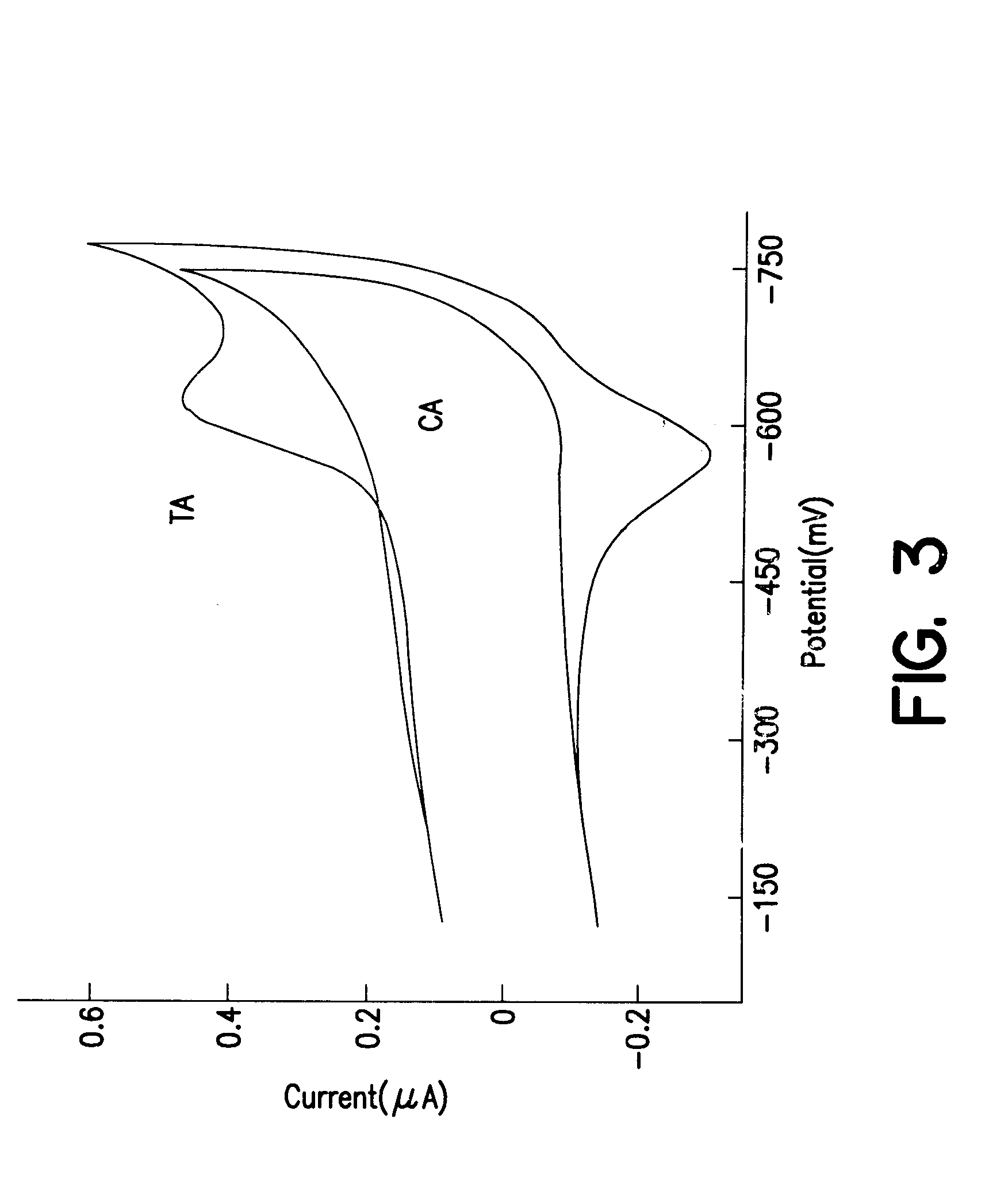
Moving the guanine site along the duplex r...

example 2

Characterization of DNA Duplexes Modified with a Redox-Active Intercalator.

Modified duplexes were characterized by mass spectrometry, ultraviolet / visible absorption spectroscopy, and thermal denaturation experiments, all of which were consistent with a 1:1 DM-duplex stoichiometry. For example, the duplex SH--(CH.sub.2)CONH(CH.sub.2).sub.6 NHCO.sub.2 -.sup.5' ATCCTACTCATGGAC with its inosine complement modified with DM was analyzed by MALDI-TOF spectrometry. Mass-to-charge ratios (found / calc) of 5284 / (5282) (DM+SH strand), 4541 / (4540) (complement), and 4742 / (4742) (SH strand) were detected. These values correspond to the calculated masses for fragments expected from this duplex. UV-visible absorption spectroscopy also revealed a 1:1 duplex / DM stoichiometry based upon comparison of the duplex absorbance at 260 nm (M=14.9.times.10.sup.3 M.sup.-1 cm.sup.-1) and the absorbance of intercalated DM at 480 nm (M=5.1.times.10.sup.3 M.sup.-1 cm.sup.-1). In the presence of 100 mM phosphate, 100...

example 3

Preparation of Gold Electrodes Derivatized with DNA Duplexes.

Electrodes were conveniently prepared by modifying gold surfaces with 15 base-pair DNA duplexes derivatized at the 5' end with a thiol-terminated alkane chain. Bulk gold electrodes were polished successively with 0.3-and 0.5-.mu.M alumina (Buhler), sonicated for 30 min, and etched in 1.0 M sulfuric acid. Au(111) surfaces were prepared by vapor deposition onto mica or glass (Widrig, 1991; Zei, 1983). Electrodes were then modified by incubation in 0.1 mM solutions of derivatized DNA duplexes in 5 mM phosphate / 50 mM NaCl (pH 7) for 12-48 h at ambient temperature. Modified electrodes were rinsed in buffer prior to use.

Before deposition of the duplexes onto the gold surfaces, the presence of the free thiols was confirmed using a spectroscopic assay based on dithionitrobenzene (Riddles, 1979). Subsequently, the samples were deposited onto the gold surfaces for 12-24 h.

Electrochemical assays, radioactive tagging experiments, and ...


PUM

PropertyMeasurementUnit
diameteraaaaaaaaaa
pHaaaaaaaaaa
temperatureaaaaaaaaaa

Abstract

Compositions and methods for electrochemical detection and localization of genetic point mutations, common DNA lesions and other base-stacking perturbations within oligonucleotide duplexes adsorbed onto electrodes and their use in biosensing technologies are described. An intercalative, redox-active moiety (such as an intercalator or nucleic acid-binding protein) is adhered and / or crosslinked to immobilized DNA duplexes at different separations from an electrode and probed electrochemically in the presence or absence of a non-intercalative, redox-active moiety. Interruptions in DNA-mediated electron-transfer caused by base-stacking perturbations, such as mutations or binding of a protein to its recognition site are reflected in a difference in electrical current, charge and / or potential.

Description

The present invention relates to the detection and localization of base-pair mismatches and other perturbations in base-stacking within an oligonucleotide duplex.DESCRIPTION OF RELATED ARTIt is now well known that mutations in DNA can lead to severe consequences in metabolic functions (e.g., regulation of gene expression, modulation of protein production) which ultimately are expressed in a variety of diseases. For example, a significant number of human cancers are characterized by a single base mutation in one of the three ras genes (Bos, 1989). In order to unravel the genetic components of such diseases, it is of utmost importance to develop DNA sensors that are capable of detecting single-base mismatches rapidly and efficiently and to establish routine screening of disease-related genetic mutations based on such sensors (Skogerboe, 1993; Southern, 1996; Chee, 1996; Eng, 1997).Various methods that have been developed for the detection of differences between DNA sequences rely on h...

Claims


Application Information

Patent Timeline
Patent Type & Authority Patents(United States)
IPC IPC(8): C12Q1/68C12N15/09C12N15/11G01N33/53G01NG01N27/327G01N27/416
CPCB82Y30/00C12Q1/6825C12Q1/6827C12Q1/6837C12Q2565/607C12Q2563/113C12Q2563/173
Inventor BARTON, JACQUELINE K.BOON, ELIZABETH M.KELLEY, SHANA O.HILL, MICHAEL G.
Owner CALIFORNIA INST OF TECH
Why Eureka
Try Eureka