Treatment apparatus for endoscope

a treatment apparatus and endoscope technology, applied in the field of endoscope treatment apparatus, can solve the problems of long treatment time, difficult observation of the incision site, and inconvenient use,

Inactive Publication Date: 2004-02-10
OLYMPUS CORP
View PDF9 Cites 211 Cited by
  • Summary
  • Abstract
  • Description
  • Claims
  • Application Information

AI Technical Summary

Problems solved by technology

The incision therefore has to be repeated many times in order to resect the mucosa in a broad range, and this is troublesome.
Moreover, since smoke or steam is generated during the incising of the tissue of a mucosa lower layer, it is difficult to observe the part to be incised because of the smoke or steam.
Additionally, to secure a field of view every incision, the smoke or steam has to be removed by an air supply and sucking operation of the endoscope, and the treatment time lengthens.
For this, complicated operations such as coupled operations for bending the endoscope, elevating the forceps, and moving the incising instrument forwards and backwards have to be carried out, these operations are laborious and require cautiousness, and the operator has to sustain concentration for a long time.
Moreover, since the mucosa is turned over, it is difficult to see the range of the mucosa lower layer to be cut.
Therefore, it is necessary to return the mucosa to its original position and to confirm the affected area, and this is laborious and troublesome.
However, an operation for incising only the mucosa with the incising instruments

Method used


Image

  • Treatment apparatus for endoscope
  • Treatment apparatus for endoscope
  • Treatment apparatus for endoscope

Examples


first embodiment

(Twenty-First Embodiment)

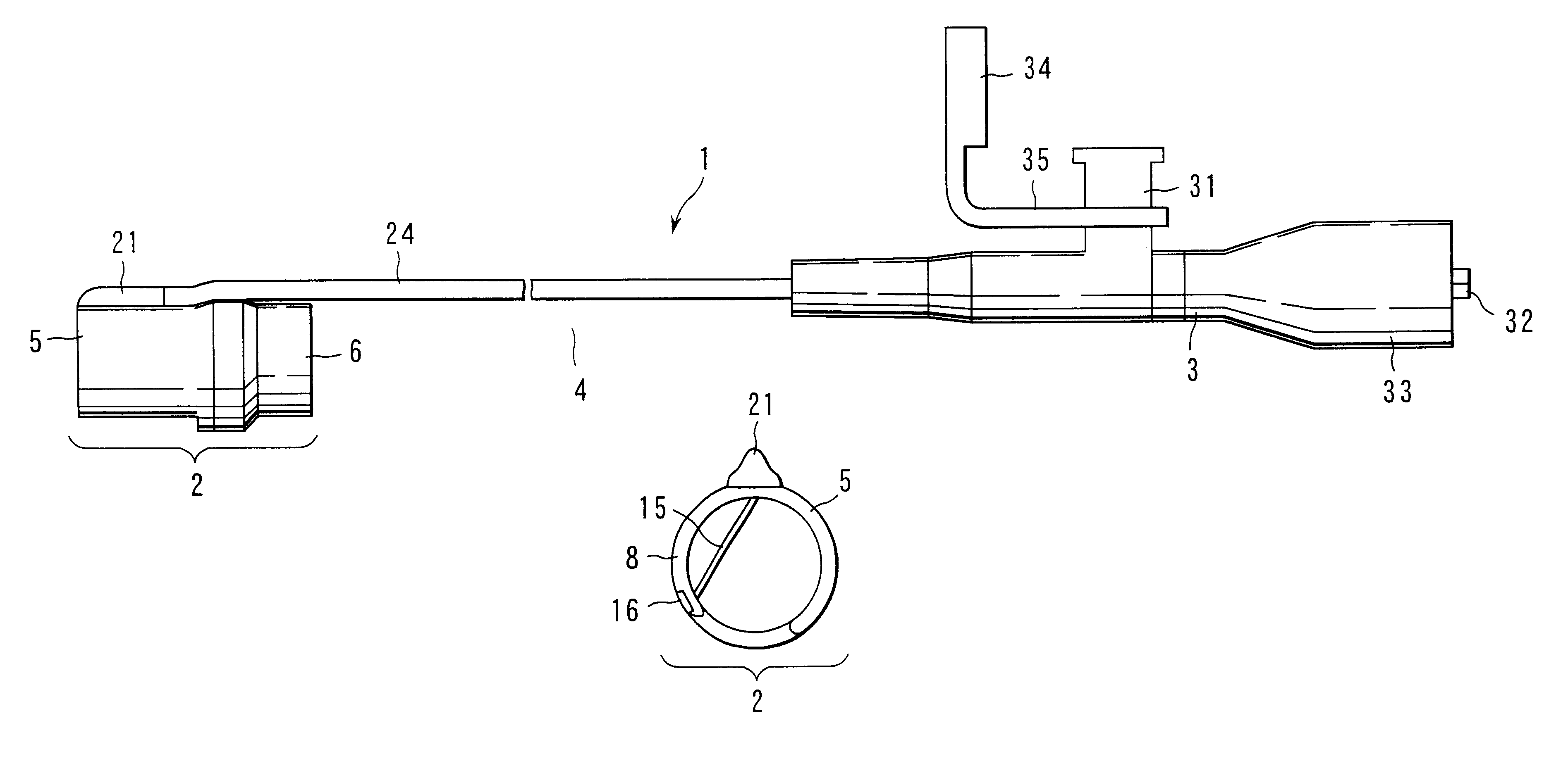
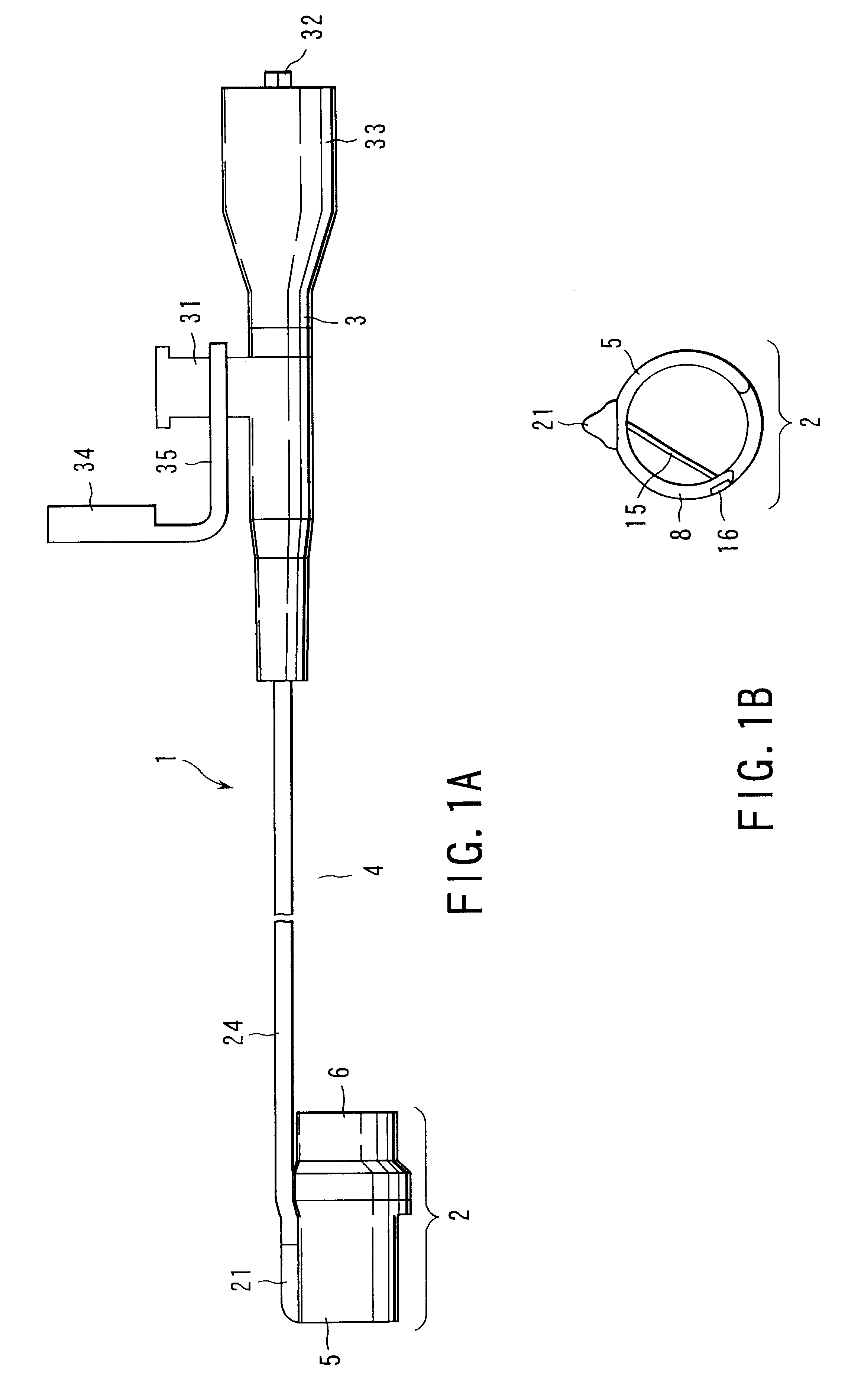
FIG. 78 shows the treatment apparatus for the endoscope according to a twenty-first embodiment.

In the twenty-first embodiment, the inner cylinder 202 of the twentieth embodiment is omitted, and instead, a cylindrical hood 260 having an attachable / detachable inner diameter and formed of a flexible and transparent resin superior in heat resistance is disposed as a tubular member in the tip-end portion 12 of the endoscope 10. The hood 260 includes the sharp edge 241, side aperture 242, incising instrument holding portion 243 and wire 247 similarly as the twentieth embodiment.

Here, the guide tube 203 does not have an inner / outer double cylinder structure, and has a single structure. The middle hole diameter of the rubber seal of the cap disposed on the rear end is slightly smaller than the outer diameter of the inserting portion 11 of the endoscope 10. Moreover, the high-frequency cable 233 is taped and fixed at several positions on the inserting portion 11 of t...

second embodiment

(Twenty-Second Embodiment)

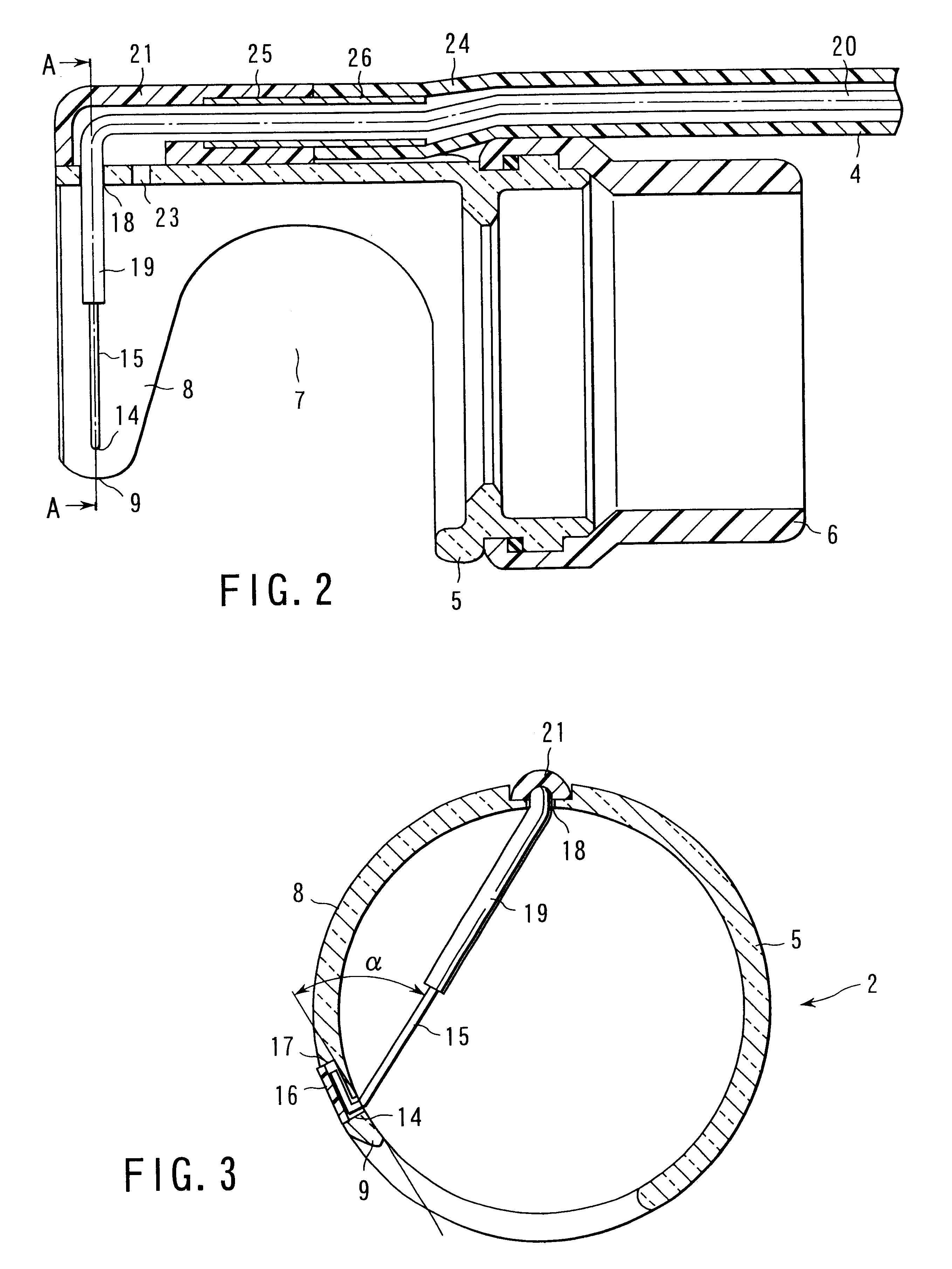
FIGS. 79A, 79B and 80 show the treatment apparatus for the endoscope according to a twenty-second embodiment.

In the twenty-second embodiment, similarly as the twenty-first embodiment, the hood 260 is used, but the side aperture 242 is formed in the hood 260 to extend in a broad range to the oblique front lower half of the hood 260. Moreover, the incising instrument holding portion 243 is disposed to project to the vicinity of the middle portion from the rear end of the side aperture 242 along the axial direction of the hood 260.

Here, the incising instrument holding portion 243 is formed in the shape of a hollow pipe, and the incising instrument introducing portion 245 formed by the tip-end portion of the incising instrument holding portion 243 is formed in a bullet shape.

Moreover, a hole 261 is made in the middle of the incising instrument introducing portion 245 of the incising instrument holding portion 243, and an elongate circular slit 262 is opened alo...

third embodiment

(Twenty-Third Embodiment)

FIG. 81 shows the treatment apparatus for the endoscope according to a twenty-third embodiment.

In the endoscope treatment apparatus of the twenty-third embodiment, as shown in FIG. 81, the attachable / detachable hood 260 is detachably attached to the tip-end portion 12 of the endoscope 10. The hood 260 is formed of a transparent and flexible polyurethane resin, and the tip-end portion thereof has a bullet shape. The side aperture 242 of the hood 260 is largely opened substantially over a 2 / 3 circumference of the hood 260. In the aperture, two incising instrument holding portions 243 are formed integrally with the hood 260, project in the axial direction and are disposed in parallel to each other. An interval of about 90 degrees for the circumferential angle is made between two incising instrument holding portions 243. On a side portion of the side aperture 242 positioned between two incising instrument holding portions 243, the sharp edge 241 is obliquely for...


PUM


Abstract

A treatment apparatus to treat a tissue in a body cavity is disclosed. The apparatus comprises a base disposed in the vicinity of a tip-end portion of an endoscope, and an in-tissue inserting portion including a base end supported by the base and a tip end insertable in the tissue in the body cavity in a direction substantially parallel to the surface of the tissue, and extending to the tip end from the base end in a tapered shape. The in-tissue inserting portion has an inner side disposed in the vicinity of the surface of the tissue when inserted into the tissue. The apparatus further comprises a high-frequency electrode, for treating the tissue, supported by the base and disposed in the vicinity of the inner side of the in-tissue inserting portion, and a cable which supplies power to the high-frequency electrode from a high-frequency power source outside the body.

Description

1. Field of the InventionThe present invention relates to a treatment apparatus for an endoscope, which is used in combination with the endoscope, and which is used in treatments such as resecting of a mucosa.2. Description of the Related ArtThere has heretofore been a strip biopsy using a grip forceps and a snare as a method of resecting an affected area in a mucosa in an endoscopic manner. One example of a method by the strip biopsy comprises: incising a part swollen in a bump shape; picking up or turning over the incised mucosa with the grip forceps introduced through a treatment instrument insertion passage (channel) of the endoscope so that a boundary tissue can be observed with the endoscope; and bringing a high-frequency or a high-energy incising instrument such as a laser scalpel introduced through the treatment instrument insertion passage of the endoscope onto the tissue and incising the tissue.In this method, the range from which the mucosa is stripped with one stroke of ...

Claims


Application Information

Patent Timeline
IPC IPC(8): A61B18/14A61B17/34A61B18/00A61B1/00A61B17/32A61B18/12
CPCA61B18/1492A61B17/3478A61B18/1482A61B2017/00269A61B2018/00601A61B2018/00982A61B2018/1407A61B2218/002A61B2017/00296A61B1/00089A61B1/00091
Inventor ARAI, KEIICHIWATANABE, HIROYOSHISUZUKI, KEITASUGI, YOSHIHIKOKAWASHIMA, KOICHIISHIKAWA, MASAHIRO
Owner OLYMPUS CORP
Why Eureka
Try Eureka