Centroid apparatus and method for sub-pixel X-ray image resolution

a technology of x-ray image resolution and centroids, which is applied in the field of x-ray imaging apparatuses, can solve the problems of limited x-ray data resolution, adverse effects on image resolution of resultant x-ray images, and limits the resolution of x-ray detectors, so as to reduce the negative effect of light spread in the scintillator on image resolution, reduce the effect of x-ray detection efficiency and reduced image resolution

Inactive Publication Date: 2005-01-25
GE MEDICAL SYST GLOBAL TECH CO LLC +1
View PDF4 Cites 2 Cited by
  • Summary
  • Abstract
  • Description
  • Claims
  • Application Information

AI Technical Summary

Benefits of technology

The electrical signals from the array of charge collection electrodes are fed to a signal processor. The signal processor analyzes the electrical signals and defines a two dimensional matrix of the charge collection electrodes in the two dimensional array. Preferably a square matrix is defined that is centered about the charge collection electrode that produced the electrical signal indicating the greatest number of photoelectron strikes. The analysis of th...

Problems solved by technology

The resolution of the resultant X-ray image was adversely affected by the diversion, or spreading, of the light within the scintillator.
The resolution of the resultant X-ray data is limited by the pitch, or spacing, of the charge collection electrodes.
Thus, the ability to physically construct the electro...

Method used


Image

  • Centroid apparatus and method for sub-pixel X-ray image resolution
  • Centroid apparatus and method for sub-pixel X-ray image resolution
  • Centroid apparatus and method for sub-pixel X-ray image resolution

Examples


Embodiment Construction

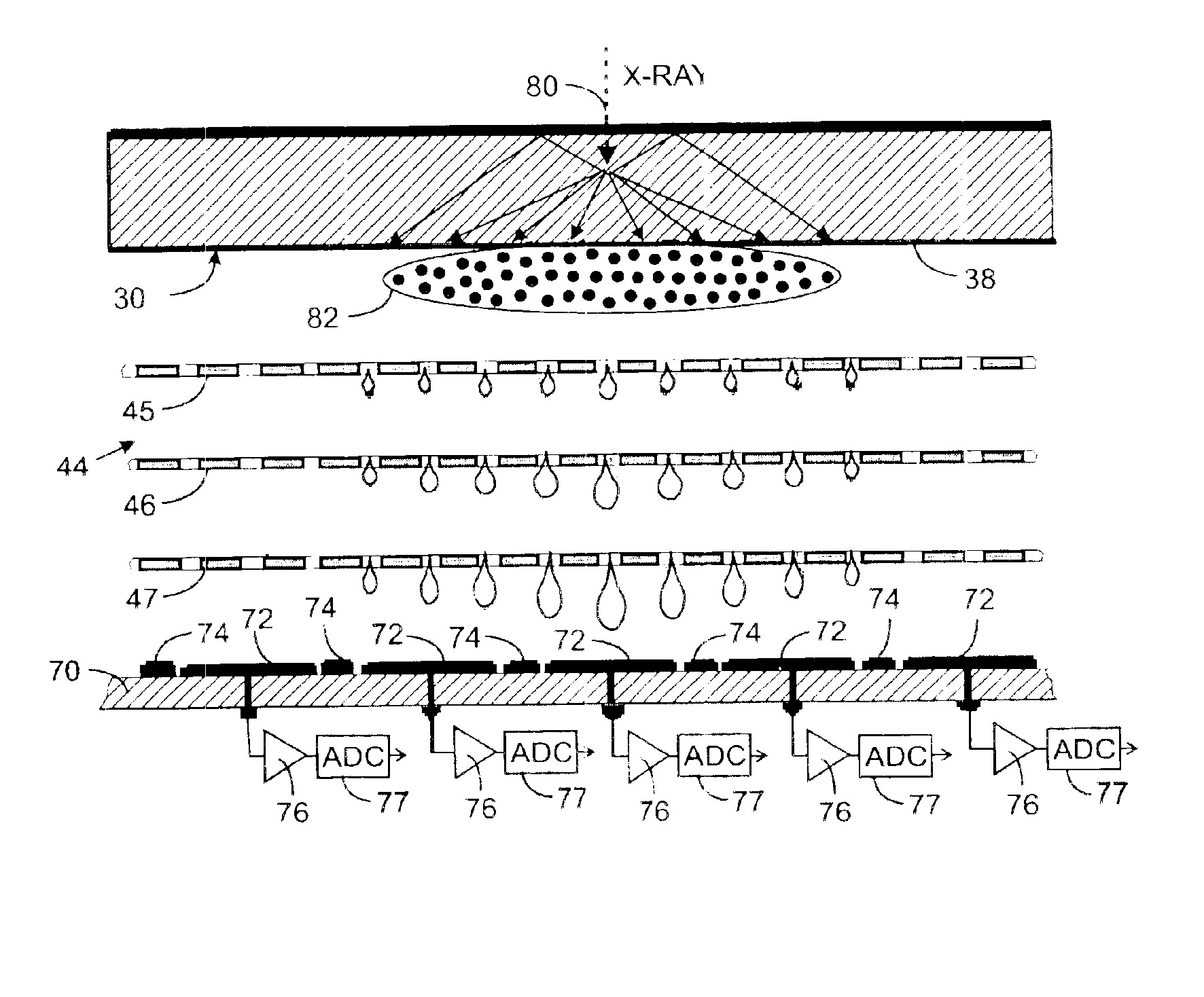
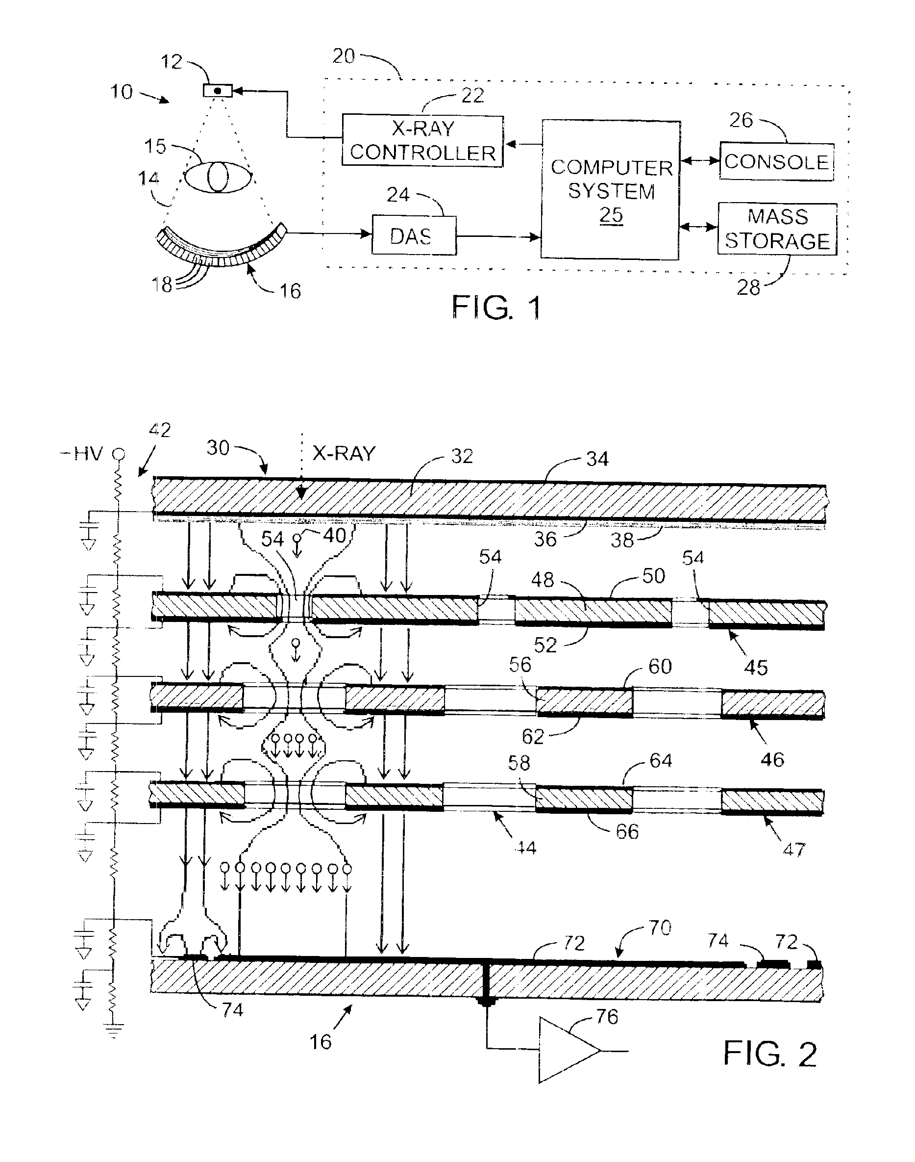
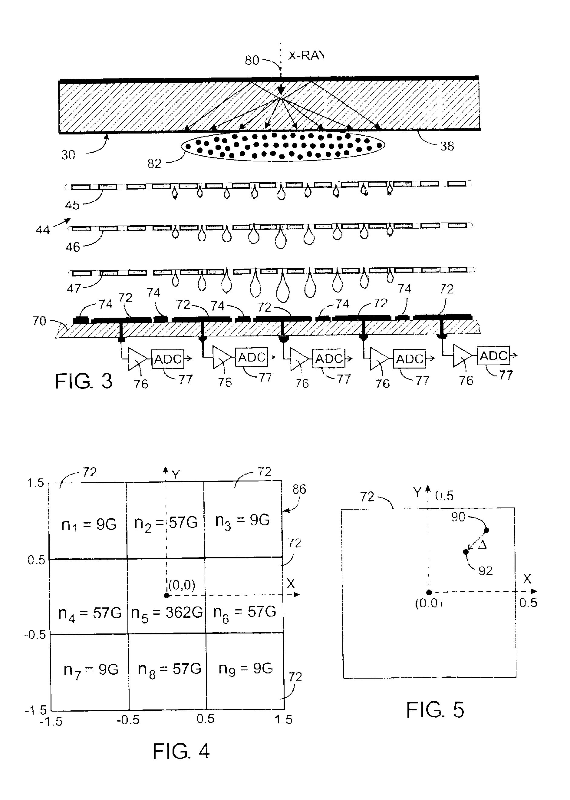
With initial reference to FIG. 1, an X-ray imaging system 10, such as used for medical imaging, has an X-ray source 12 that projects a cone beam of X-rays 14 toward a detector array 16 on the opposite side of the medical patient being imaged. The detector array 16 is formed by two dimensional array of a plurality of detector elements 18 which together sense the projected X-rays that pass through a patient 15. The impact of an X-ray photon on the detector array 16 is known as a photon event and produces electrical signals from several of the detector elements 18 as will be described. The detector array 16 has circuits which digitize the detector element signals.

Operation of the X-ray source 12 is governed by a control and image processing system 20 which includes an X-ray controller 22 that provides power and timing signals to the X-ray source 12. A data acquisition system (DAS) 24 samples data produced by detector elements 18. Operation of the X-ray controller 22 and the data acquis...


PUM


Abstract

An apparatus for detecting X-rays comprises a scintillator which emits a plurality of photoelectrons upon being impacted by an X-ray photon. The photoelectrons are amplified in a gas electron multiplier and the resultant photoelectrons are accumulated on a two dimensional array of charge collection electrodes. Electrical signals are produced which indicate the quantity of photoelectrons which strike each charge collection electrode. A processor determines a location of the X-ray photon strike by analyzing the spatial distribution of the photoelectrons accumulated by the array of charge collection electrodes. The intensity of the X-ray photon is determined from the number of accumulated photoelectrons.

Description

CROSS-REFERENCE TO RELATED APPLICATIONSNot applicable.STATEMENT REGARDING FEDERALLY SPONSORED RESEARCH OR DEVELOPMENTNot applicable.BACKGROUND OF THE INVENTION1. Field of the InventionThe present invention relates to X-ray imaging apparatus; and more particularly to X-ray detectors which produce electrical image signal in such apparatus.2. Description of the Related ArtConventional X-ray imaging equipment includes a source for projecting a beam of X-rays through an object being imaged, such as a medical patient. The portion of the beam which passes through the patient impinges upon an X-ray detector which converts the X-rays attenuated by the patient into photons which then are converted into an electric image signal. One type of X-ray detector has a combination of a scintillator in front of a two dimensional array of photodetectors. Each photodetector integrates the energy of the impacting X-ray photons over the period of X-ray exposure time to produce a signal that is proportional...

Claims


Application Information

Patent Timeline
IPC IPC(8): G01T1/20G01T1/00G01T1/29H01J37/244H01J47/02H01J47/00H01J43/00H05G1/64H05G1/00H04N5/321H04N5/325H01J31/49
CPCH01J47/02
Inventor MAOLINBAY, MANATGRANFORS, PAUL R.
Owner GE MEDICAL SYST GLOBAL TECH CO LLC
Why Eureka
Try Eureka