Retinal cell lines with extended life-span and their applications

a technology of retinal cells and life-span, applied in the field of new cells with extended life-span of retinal origin, can solve problems such as rejection reactions, and achieve the effect of prolonging life-span and being better able to meet practical needs

Inactive Publication Date: 2005-04-12
NEUROTECH USA
View PDF7 Cites 10 Cited by
  • Summary
  • Abstract
  • Description
  • Claims
  • Application Information

AI Technical Summary

Benefits of technology

The invention provides lines of cells with extended life-span derived from primary cultures of the endothelium of the blood-retina barrier and the retinal pigment epithelium of mammals. These lines of cell are better capable of meeting practical needs, especially in that all the lines obtained are stable and have most of the characteristics of the differentiated cells.
The invention also provides cell lines derived from the lines with extended life-span as defined above (“vector lines”), characterized(a) in that they comprise at least one cell line as defined above, associated with an expression vector comprising a sequence coding for a polypeptide, a protein, or a viral vector, optionally associated with at least one selection gene, and optionally associated with at least one marker gene, and(b) in that they are capable, in vivo, of integrating into the retina and especially the subretinal space of a host mammal, preventing the loss of photoreceptors and producing the peptide, the protein or the viral vector.

Problems solved by technology

For in vivo transfer, the use of primary nerve tissues of fetal origin for cellular transplantation in human therapy gives rise to numerous ethical and practical problems.
Such cells can modify the normal function of the nervous system and cause a rejection reaction.

Method used


Image

  • Retinal cell lines with extended life-span and their applications
  • Retinal cell lines with extended life-span and their applications
  • Retinal cell lines with extended life-span and their applications

Examples


example 1

Methods Used to Characterize the Cells of the Invention

(a) Isolation and culture of rat aortic endothelial cells. The aortic endothelium was isolated by the method described by McGuire et al., 57 Lab. Invest. 94-105 (1987). The rat aorta was removed by dissection and cut into small pieces (2-5 mm), which were placed on 24-well plates coated with collagen and containing an endothelial cell culture medium, in such a way that the luminal face of the pieces of aorta was in contact with the collagen. The RPMI culture medium was supplemented with 20% fetal calf serum, 7.5 μg / ml endothelial cell growth supplement, 80 μg / ml heparin, 2 mM glutamine, 0.5 μg / ml vitamin C, 100 U / ml penicillin, and 100 μg / ml streptomycin.

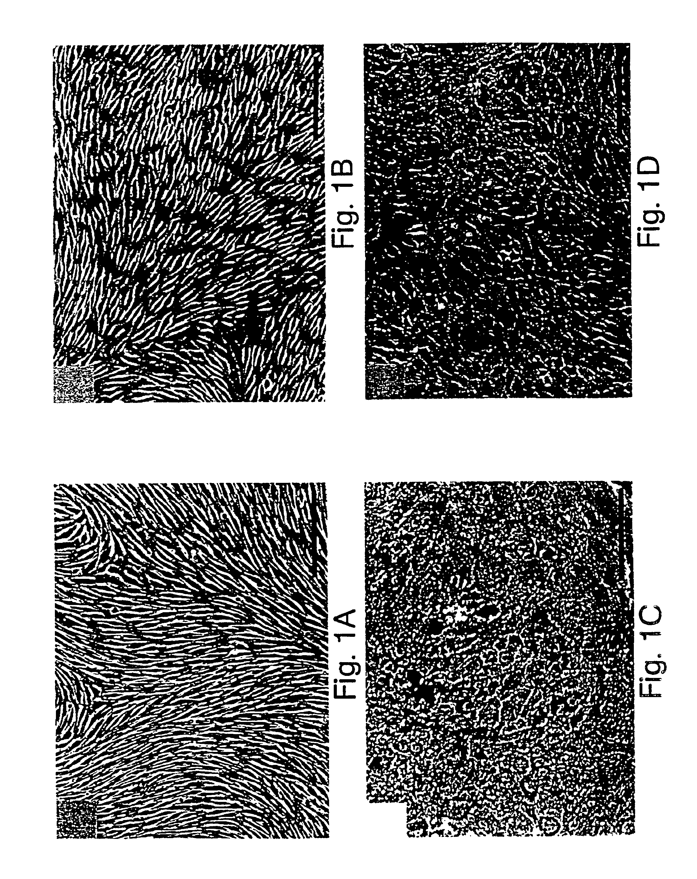
After 3 days, the explants were removed and the adhering cells were proliferated to the point of confluence. At confluence, the cells had a pavement morphology characteristic of the endothelial vessels, expressed Von Willebrand's factor, and proliferated in a medium containing D...

example 2

Preparation of a Line According to the Invention

Rat Retinal Endothelial Cells

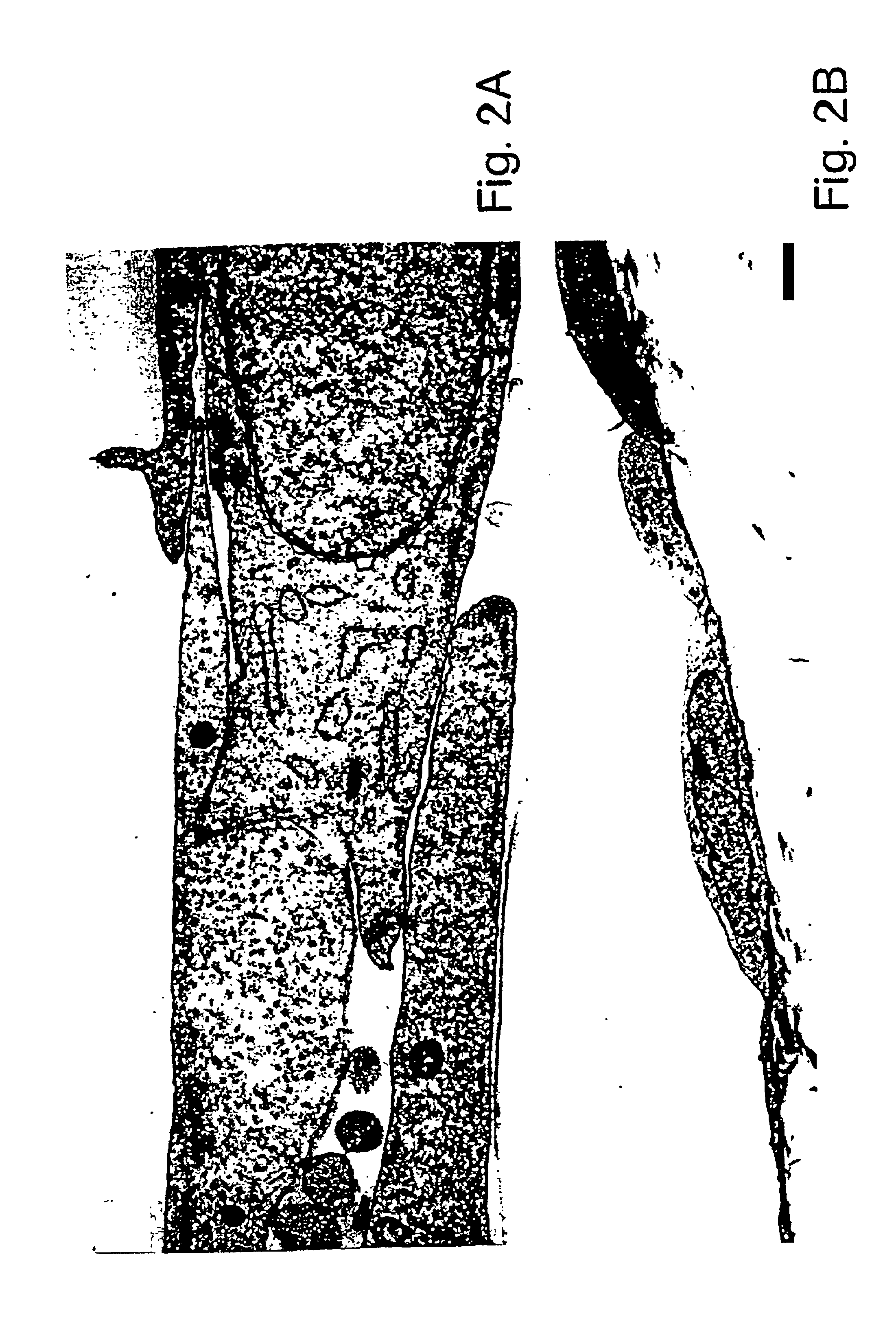
(a) Isolation and culture of the retinal endothelial cells. Endothelial cells were derived from 4- to 6-week-old female Lewis rats free of pathogens. The retinal cells were isolated and cultivated by the methods described by Greenwood, 39 J. Neuroimmunol. 123-132 (1992) and Abbott et al., 103 Cell Sci. 23-37 (1992). These techniques produced primary cultures with a purity in excess of 95%. The rat retinas were dispersed by enzymatic digestion, the fragments of microvessels were separated from the cells themselves by centrifugation and the cells were washed and cultured in flasks coated with collagen. The growth medium consisted of Ham's F-10 medium (Sigma, St. Louis, Mo.) supplemented with 17.5% serum (Advanced Protein Products Ltd.); 7.5 μg / ml of endothelial cell growth supplement (Advanced Protein Products Ltd.); 80 μg / ml of heparin, 2 mM glutamine; 0.5 μg / ml of vitamin C; 100 U / ml of penicillin; and 100 ...

example 3

Preparation of a Line According to the Invention

Rat Retinal Epithelial Cells

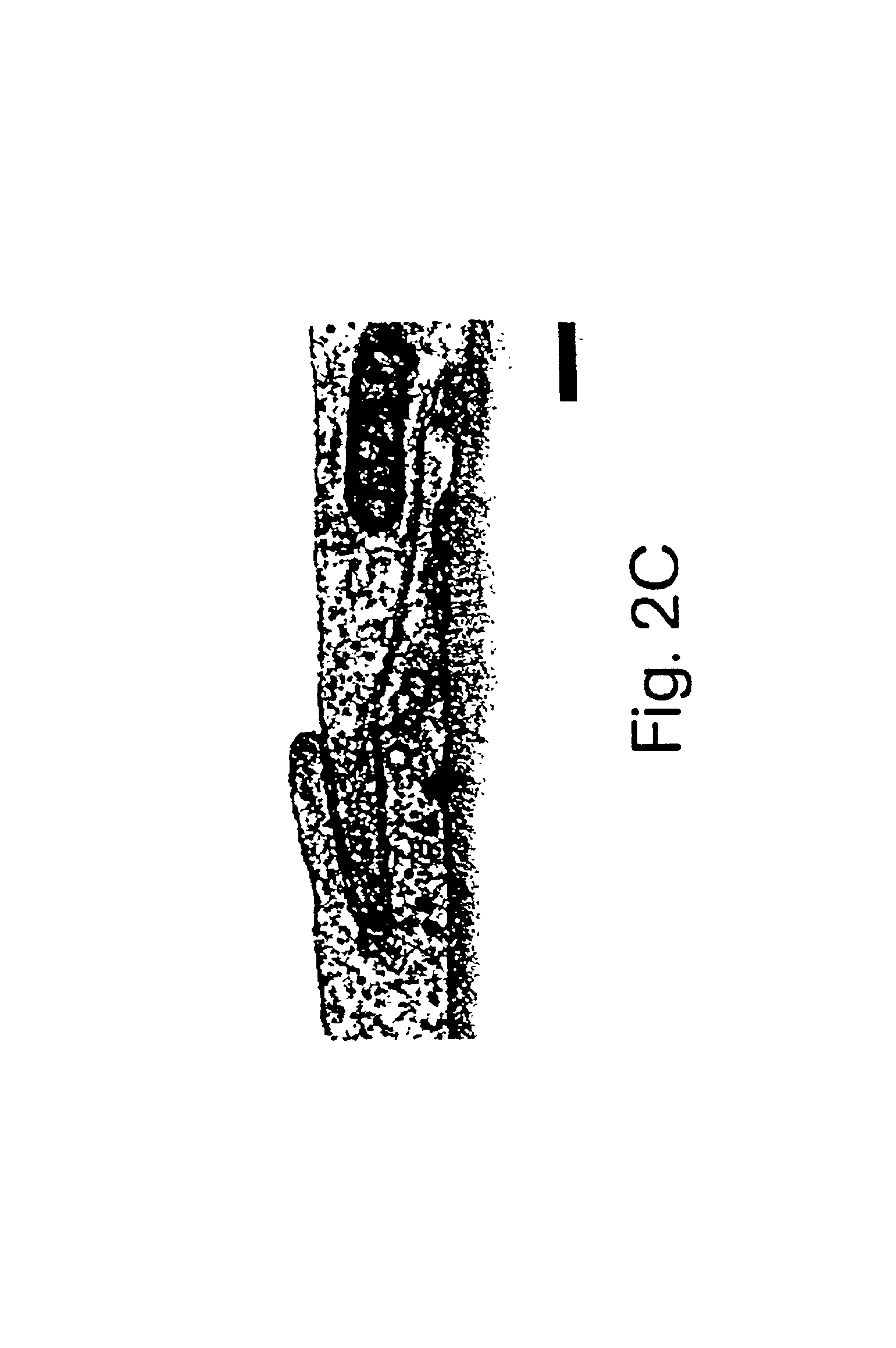
(a) Isolation and culture of the retinal pigment epithelial cells. The rat retinal pigment epithelial cells were isolated from 6-day to 8-day old PVG rats according to the method of Chang et al., 10 Curr. Eye Res. 1081-1086 (1991). The eyes were removed and the intact eyeballs were digested with 2% dispase for 30 min. The eyes were then dissected for removal of the cornea and the vitreous body. The dissected retina was then isolated and incubated for 15 min in a culture medium. After incubation, layers of retinal pigment epithelial cells were separated from the neuroretina and treated with trypsin to produce a cellular suspension. The cells were plated in tissue culture flasks and cultivated to the point of semiconfluence. The culture medium consisted of Ham's F-10 medium supplemented with 20% fetal calf serum, 20 mM HEPES, 7.5% sodium bicarbonate, 2 mM glutamine, 100 U / ml of penicillin and 100 μg / ml of stre...


PUM

PropertyMeasurementUnit
timeaaaaaaaaaa
pHaaaaaaaaaa
pHaaaaaaaaaa

Abstract

The invention features retina-derived (retinal endothelial or retinal epithelial pigment) cell lines with extended life-span and capable of being implanted in the retina and of carrying a therapeutic substance to the eye and to the central nervous system. Such lines can also be used as a model for studying blood central nervous system interfaces. These lines are derived from primary retinal cultures selected from the group consisting of primary retinal endothelial cells and primary retinal epithelial cells, comprise a polynucleotide containing an oncogene, which polynucleotide is optionally associated with at least one selection gene, and have the morphological characteristics and at least the expression characteristics of the surface antigens of corresponding primary cultures.

Description

TECHNICAL FIELD OF THE INVENTIONThe present invention relates to novel cell lines with extended life-span of retinal origin (retinal endothelial and retinal pigment epithelial origin) that are capable of being implanted in the retina and of conveying a substance of therapeutic interest into the eye or the central nervous system.BACKGROUND OF THE INVENTIONBoth the blood-brain barrier and the blood-retina barrier are important in controlling the passage of substances to and from the neural parenchyma, especially in the maintenance of homeostasis.In the retina, the blood-retina barrier is composed of two different types of anatomically separate cells. The retinal vascular endothelium, which supplies the anterior portion of the retina, is currently considered to have an identical structure to the cerebral endothelium (Towler et al., 480 J. Physiol. 10-11P (1994)), whereas the cells of the retinal pigment epithelium cover the permeable vessels of the choroidal circulation and form the po...

Claims


Application Information

Patent Timeline
Patent Type & Authority Patents(United States)
IPC IPC(8): C12N5/06A61K35/12A61K35/44C12N15/09A61P27/02A61P29/00A61P35/00C12N5/071C12N5/10C12P21/02
CPCC12N5/0621A61K35/12C12N2510/04C12N2510/02C12N2503/00A61P27/02A61P29/00A61P35/00
Inventor GREENWOOD, JOHNADAMSON, PETERLUND, RAYMONDTAO, WENG
Owner NEUROTECH USA
Why Eureka
Try Eureka