Method, system and computer readable medium for an intelligent search workstation for computer assisted interpretation of medical images

a workstation and medical image technology, applied in the field of methods, can solve the problems of estimated 46,000 deaths per year and considerable misclassification of lesions

Inactive Publication Date: 2005-05-31
ARCH DEVMENT
View PDF26 Cites 68 Cited by
  • Summary
  • Abstract
  • Description
  • Claims
  • Application Information

AI Technical Summary

Problems solved by technology

Breast cancer is a leading cause of death in women, causing an estimated 46,000 deaths per year.
Although general rules for the differentiation between benign and malignant mammographically identified breast lesions exist, considerable misclassification of lesions occurs with the current methods.

Method used


Image

  • Method, system and computer readable medium for an intelligent search workstation for computer assisted interpretation of medical images
  • Method, system and computer readable medium for an intelligent search workstation for computer assisted interpretation of medical images
  • Method, system and computer readable medium for an intelligent search workstation for computer assisted interpretation of medical images

Examples


Embodiment Construction

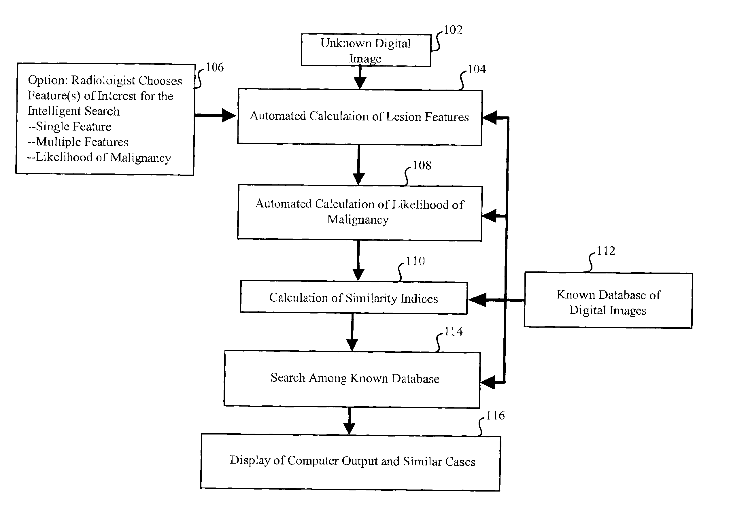
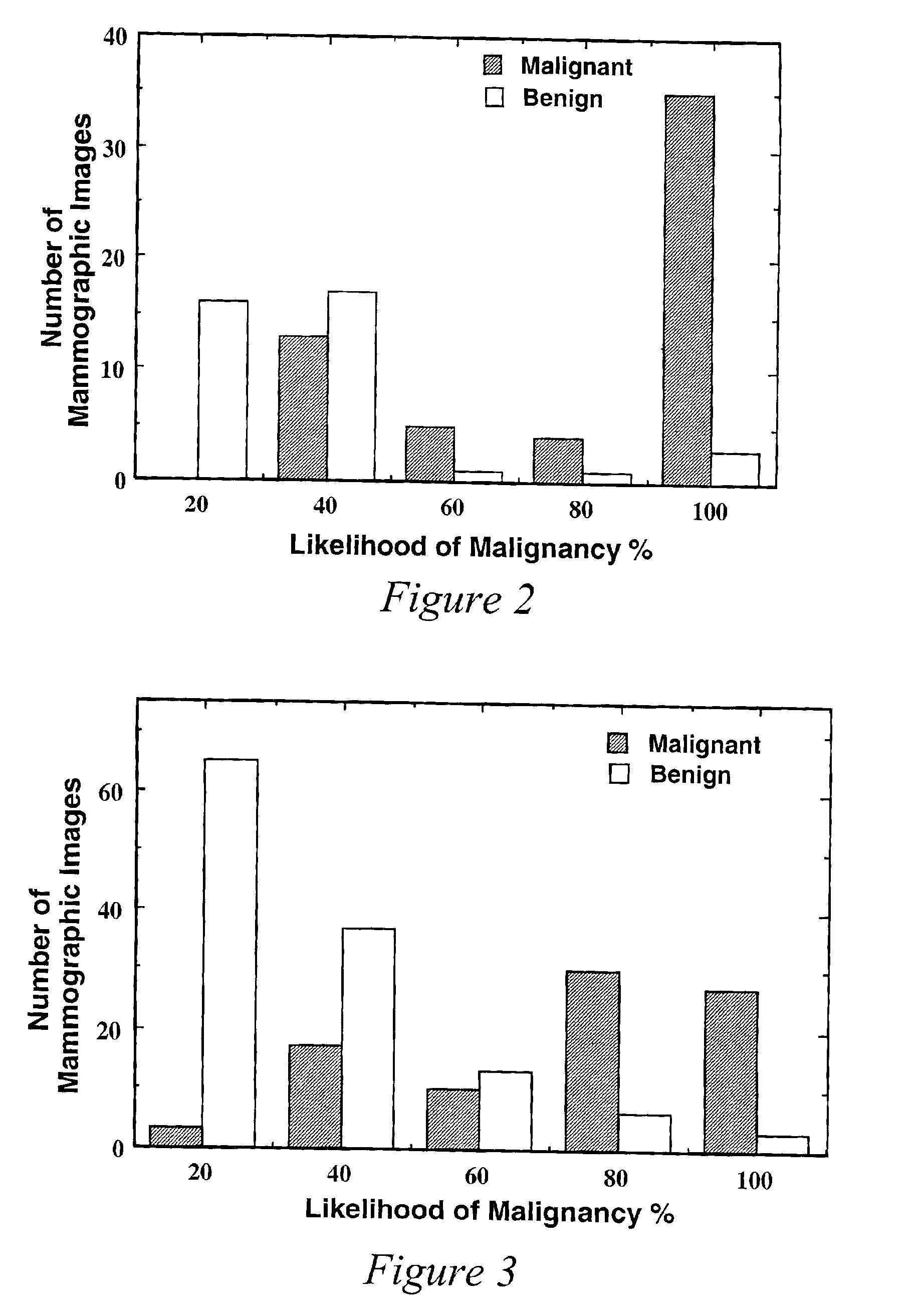
[0024]The inventors are investigating the potential usefulness of computer-aided diagnosis as an aid to radiologists in the characterization and classification of mass lesions in mammography. Observer studies show that such a system can aid in increasing the diagnostic accuracy of radiologists both in terms of sensitivity and specificity. The present mass classification method, system and computer readable medium includes three components: (1) automated segmentation of mass regions, (2) automated feature-extraction, and (3) automated classification [1-3]. The method is initially trained with 95 mammograms containing masses from 65 patients. Features related to the margin, shape, and density of each mass are extracted automatically from the image data and merged into an estimate of the likelihood of malignancy using artificial neural networks (ANNs). These features include a spiculation measure, a radial gradient index, and two density measures. The round-robin performance of the com...


PUM


Abstract

A method, system and computer readable medium for an intelligent search display into which an automated computerized image analysis has been incorporated. Upon viewing an unknown mammographic case, the display shows both the computer classification output as well as images of lesions with known diagnoses (e.g., malignant vs. benign) and similar computer-extracted features. The similarity index used in the search can be chosen by the radiologist to be based on a single feature, multiple features, or on the computer estimate of the likelihood of malignancy. Specifically the system includes the calculation of features of images in a known database, calculation of features of an unknown case, calculation of a similarity index, display of the known cases along the probability distribution curves at which the unknown case exists. Techniques include novel developments and implementations of computer-extracted features for similarity calculation and novel methods for the display of the unknown case amongst known cases with and without the computer-determined diagnoses.

Description

CROSS-REFERENCE TO RELATED APPLICATIONS[0001]This application claims priority under 35 U.S.C. § 119(e) to United States provisional patent applications Ser. No. 60 / 180,162, filed on Feb. 4, 2000 and Ser. No. 60 / 207,401, filed on May 30, 2000, the entire contents of both of which are incorporated by reference herein.BACKGROUND OF THE INVENTION[0002]1. Field of the Invention[0003]The invention relates generally to the field of computer-aided diagnosis and image display workstations. It describes a method, system and computer readable medium that employs an intelligent search workstation for the computer assisted interpretation of medical images. Upon viewing an unknown medical case, the workstation shows both computer analysis output as well as images of cases with known diagnoses (e.g., malignant vs. benign) and similar computer-extracted features. The similarity index used in the search can be chosen by the radiologist to be based on a single feature, multiple features, or on the co...

Claims


Application Information

Patent Timeline
Patent Type & Authority Patents(United States)
IPC IPC(8): G06K9/00A61B6/00G06Q50/00G06T1/00G06T7/00G16H30/40
CPCG06F19/321G06F19/345G06T7/0012G06F19/3443A61B6/502G06T2207/30068G16H50/70G16H50/20G16H30/40
Inventor GIGER, MARYELLEN L.VYBORNY, CARL J.HUO, ZHIMINLAN, LI
Owner ARCH DEVMENT
Why Eureka
Try Eureka