

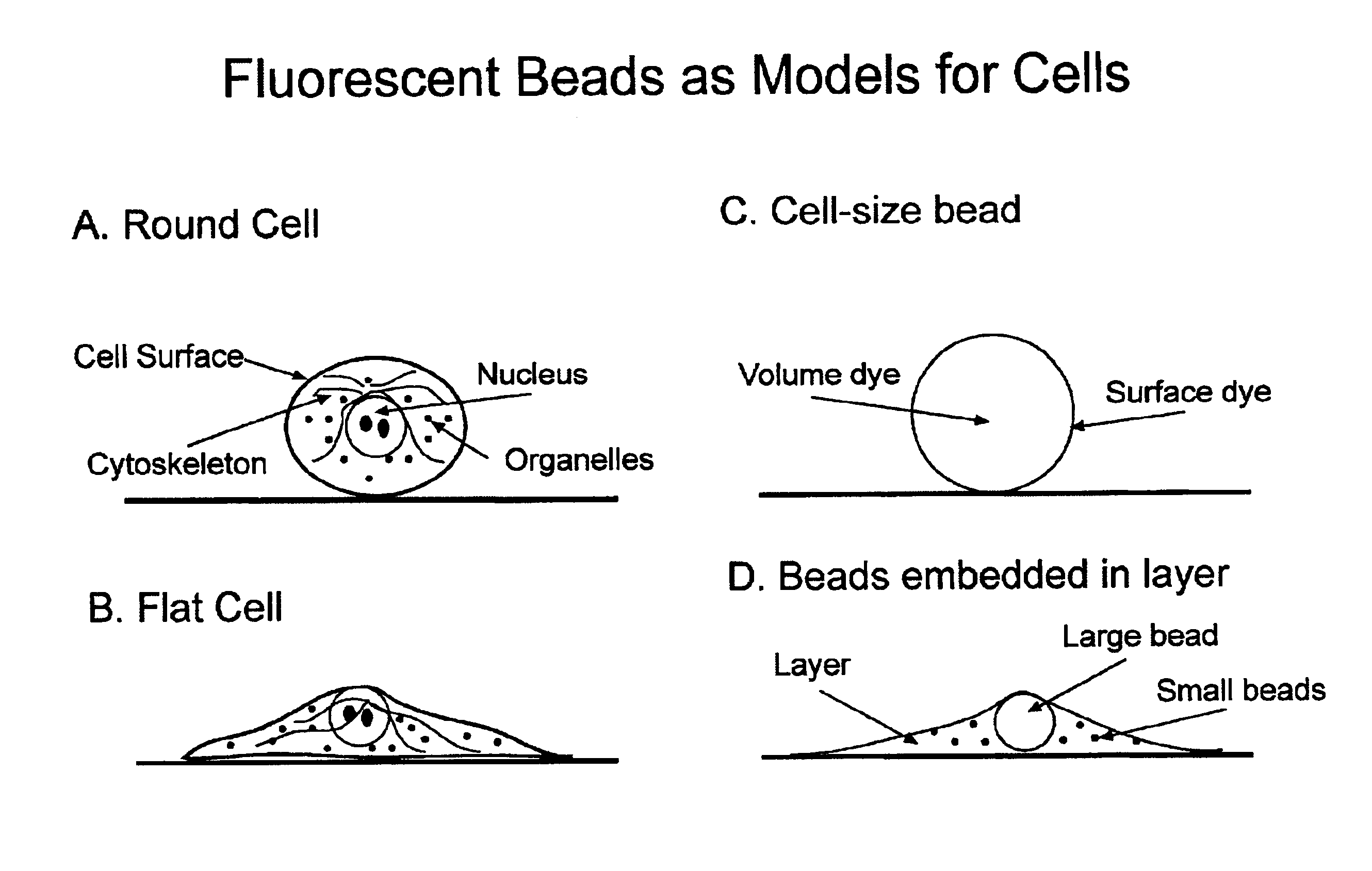
Microbead-based test plates and test methods for fluorescence imaging systems
a technology of fluorescence imaging and test plates, applied in the field of cell-based imaging, can solve the problems of not allowing calibration of imaging systems for image-based microscopic measurements, and no tools designed for diagnostics, calibration or software validation of fluorescence imaging systems that carry out image-based microscopic measurements
- Summary
- Abstract
- Description
- Claims
- Application Information
AI Technical Summary
Benefits of technology
Problems solved by technology
Method used
Image

Examples
example 4
Diagnostics, Calibration, and Validation of the Optics, Illumination, Image Analysis Architecture, and Software of a Fluorescence Imaging System
A Method to Test the Origin of Stage Coordinate System
Problem:
[0120]The location of the plate relative to the imaging system is usually not fixed. The origin of the stage relative to the imaging system can be determined using the calibration plate.
Method:
[0121]If a well of the calibration plate has beads with sufficient density, or has beads of one color and a uniform film of another color, then a method can be employed to accurately find the edges of the well, and thus infer the location of the well center relative to the stage coordinate system.
[0122]This method is only necessary as part of installation of the instrument since the location of the plate within the coordinate system of the stage is fixed and should not vary over time. One approach is as follows:
[0123](1) Allow user to move stage (via software control) until objective is unde...
example 1
Exciter A has Failing Performance and Needs to be Replaced
[0228]
TABLE 3Exciter AExciter BExciter CDichroic emitter pair XCh. 1 FailsCh. 4 PassesCh. 7 PassesDichroic emitter pair YCh. 2 FailsCh. 5 PassesCh. 8 PassesDichroic emitter pair ZCh. 3 FailsCh. 6 PassesCh. 9 Passes
example 2
Exciter A and Exciter B were Switched by Installers, and thus their Positions in the Imaging System Need to be Reversed
[0229]
TABLE 4Exciter AExciter BExciter CDichroic emitter pair XCh. 1 FailsCh. 4 FailsCh. 7 PassesDichroic emitter pair YCh. 2 FailsCh. 5 FailsCh. 8 PassesDichroic emitter pair ZCh. 3 FailsCh. 6 FailsCh. 9 Passes
PUM
| Property | Measurement | Unit |
|---|---|---|
| diameter | aaaaa | aaaaa |
| diameter | aaaaa | aaaaa |
| diameter | aaaaa | aaaaa |
Abstract
Description
Claims
Application Information