Method and apparatus for computed tomography imaging

a computed tomography and computed tomography technology, applied in the field of medical imaging arts, can solve the problems of affecting the development of new or improved pipeline features or capabilities, affecting the efficiency of computed tomography, and the hardware pipeline paradigm does not readily support optimal allocation of reconstruction resources across imaging systems, so as to improve image reconstruction speed, improve flexibility in updating, and improve the effect of image processing resources

Inactive Publication Date: 2005-06-14
KONINKLIJKE PHILIPS ELECTRONICS NV
View PDF2 Cites 19 Cited by
  • Summary
  • Abstract
  • Description
  • Claims
  • Application Information

AI Technical Summary

Benefits of technology

[0015]One advantage of the present invention resides in better allocation of image processing resources for improved image reconstruction speed.
[0016]Another advantage of the present invention resides in improved flexibility in updating, upgrading, and revising imaging facility capabilities.
[0017]Another advantage of the present invention resides in improved interconnectedness of resources of an imaging facility within the facility.
[0018]Another advantage of the present invention lies in its scalable, modular design such that it is easily mapped to meet a wide range of product performance and cost objectives.
[0019]Yet another advantage of the present invention resides in flexibility with respect to hardware. The reconstruction pipeline is constructed from general purpose processors and / or computers rather than dedicated reconstruction hardware. However, the reconstruction pipeline constructed using general-purpose components also can incorporate dedicated hardware if it is available.
[0020]Still yet another advantage of the present invention lies in its modular design that encourages incremental development and upgrading of a CT or other type of imaging facility.

Problems solved by technology

However, past reconstruction methods and apparatuses have struggled to keep up with the data influx.
Development of new or improved pipeline features or capabilities is similarly hampered.
Furthermore, the hardware pipeline paradigm does not readily support optimal allocation of reconstruction resources across imaging systems.
Interconnection of reconstruction components of different CT imaging systems is difficult or impossible.
A second CT imaging system would typically be unable to access or utilize the hardware pipeline associated with a first CT imaging scanner.
Yet another disadvantage of past methods and apparatus is that alternative reconstruction algorithms often need additional or different dedicated hardware in the reconstruction pipeline.
Conversely, if the scanner is in use for imaging, the reconstruction pipeline is unavailable to other medical personnel.

Method used


Image

  • Method and apparatus for computed tomography imaging
  • Method and apparatus for computed tomography imaging
  • Method and apparatus for computed tomography imaging

Examples


Embodiment Construction

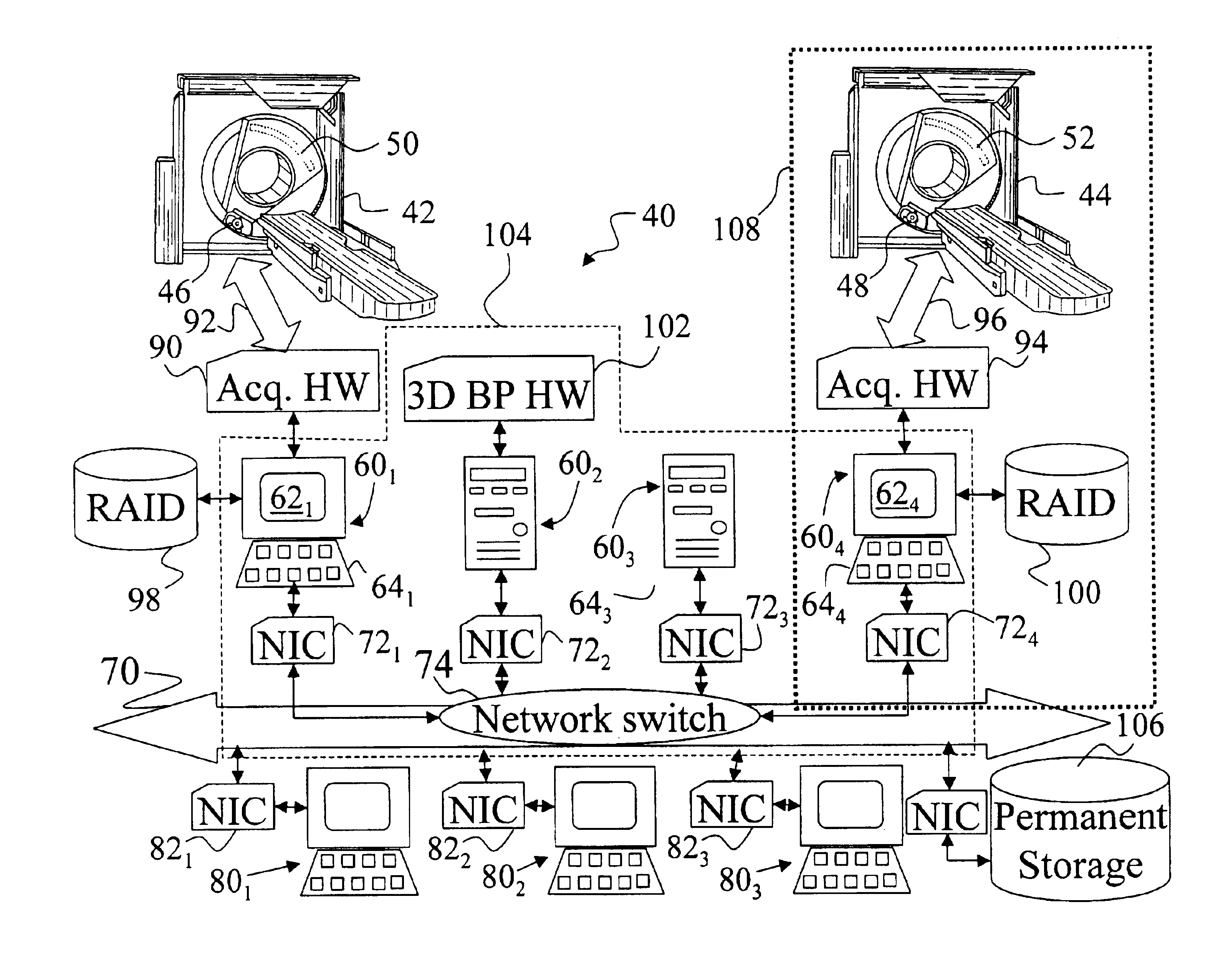
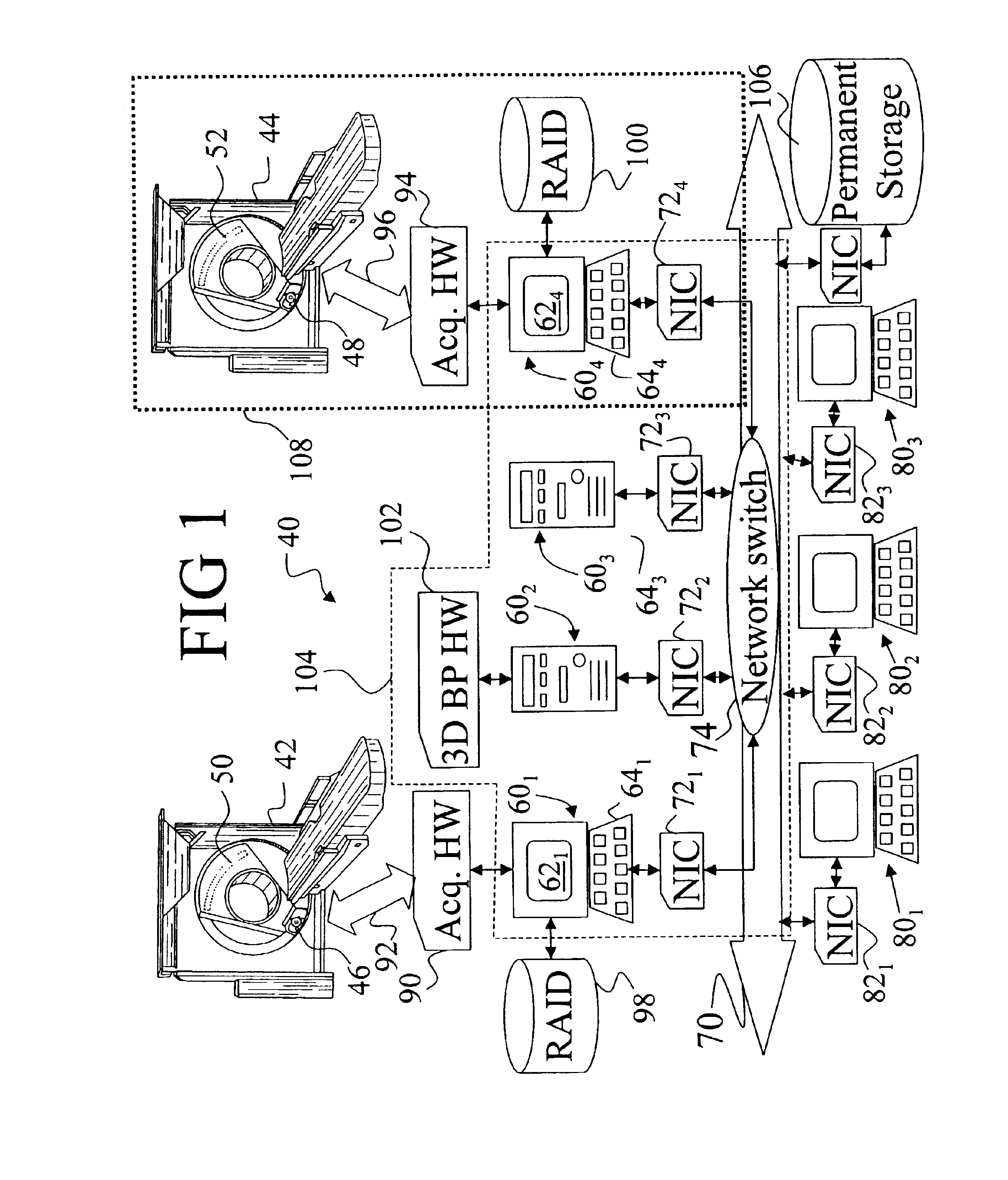
[0027]With reference to FIG. 1, an exemplary computed tomography facility 40 includes two computed tomography (CT) scanner gantries 42, 44, employing x-ray radiation sources 46, 48 and one- or two-dimensional detector arrays 50, 52. Although the two CT imaging scanners 42, 44 shown in FIG. 1 are substantially similar, the imaging scanners can also be of different configurations. One or both scanners 42, 44 can employ a conebeam helical geometry, a multi-slice wedge geometry, or other geometry. Furthermore, the facility 40 is not limited to two scanners 42, 44, but rather can include any number of scanners such as only a single CT scanner, three scanners, four scanners, and so forth.

[0028]Those skilled in the art will also recognize that the invention is not limited to computed tomography scanners employing x-ray sources, but rather is also applicable to other imaging scanners such as magnetic resonance imaging (MRI) scanners, positron emission tomography (PET) scanners, single photo...


PUM


Abstract

A computed tomography (CT) imaging apparatus (40) includes a CT imaging scanner (42) that acquires projection data. Data acquisition electronics (90) communicate with the CT scanner (42), and an acquisition computer (601) receives the projection data via the data acquisition electronics (90). A communication network (70) communicates with the acquisition computer (601) and with at least one additional computer (602, 603, 604). The at least one additional computer (602, 603, 604) cooperates with the data acquisition computer (601) and the communication network (70) to define a computational pipeline (104) that reconstructs selected projection data into a selected image representation.

Description

BACKGROUND OF THE INVENTION[0001]The present invention relates to the medical imaging arts. It particularly relates to computed tomography (CT) imaging employing a rotating x-ray source, and will be described with particular reference thereto. However, the invention will also find application in conjunction with other types of medical imaging including magnetic resonance imaging (MRI), positron emission tomography (PET), single photon emission computed tomography imaging (SPECT), diagnostic ultrasound imaging, and the like.[0002]Computed tomography (CT) imaging has application in many areas involving non-invasive examination of internal features of a subject. For example, CT is applicable in security baggage examination as well as in medical imaging. In CT imaging, an x-ray source transmits a fan or cone of x-rays into an examination region. The x-rays along each ray or path through the subject are partially absorbed. Detectors arranged across the examination region from the x-ray s...

Claims


Application Information

Patent Timeline
Patent Type & Authority Patents(United States)
IPC IPC(8): G06T11/00G01T1/161A61B5/055A61B6/03A61B8/00
CPCG06T1/20G06T11/006Y10S378/901G06T2211/428
Inventor KLING, PATRICK J.HESCHT, DONALD A.HUANG, XIANGYUMITCHKASH, GEORGE M.
Owner KONINKLIJKE PHILIPS ELECTRONICS NV
Why Eureka
Try Eureka