Multi-lumen endotracheal tube

a multi-lumen, endotracheal tube technology, applied in the field of endotracheal tubes, can solve the problems of high cost, complicated, and limit the use of optically guided intubation methods,

Inactive Publication Date: 2005-07-19
MOORE JOHNNY V
View PDF11 Cites 52 Cited by
  • Summary
  • Abstract
  • Description
  • Claims
  • Application Information

AI Technical Summary

Benefits of technology

[0013]To provide the health care practitioner the ability to monitor a patient's body core temperature, the ET tube of the present invention is provided with a temperature sensor, such as a thermistor. The temperature sensor extends into the subjacent cavity of the ausculatory receiver. The

Problems solved by technology

Merdeth also discusses the limitations of optically guided methods of intubation, citing their complexity, high cost, limited availability and limited usefulness when significant amounts of bodily fluids are present during intubation.
Accordingly, while the prior art demonstrates distinct improvements in ET tube capabilities the increasing complexity of these improvements have resulted in a concomitant increase in their cost and reduction in their availability.
Regardless of insertion order, t

Method used


Image

  • Multi-lumen endotracheal tube
  • Multi-lumen endotracheal tube
  • Multi-lumen endotracheal tube

Examples


Embodiment Construction

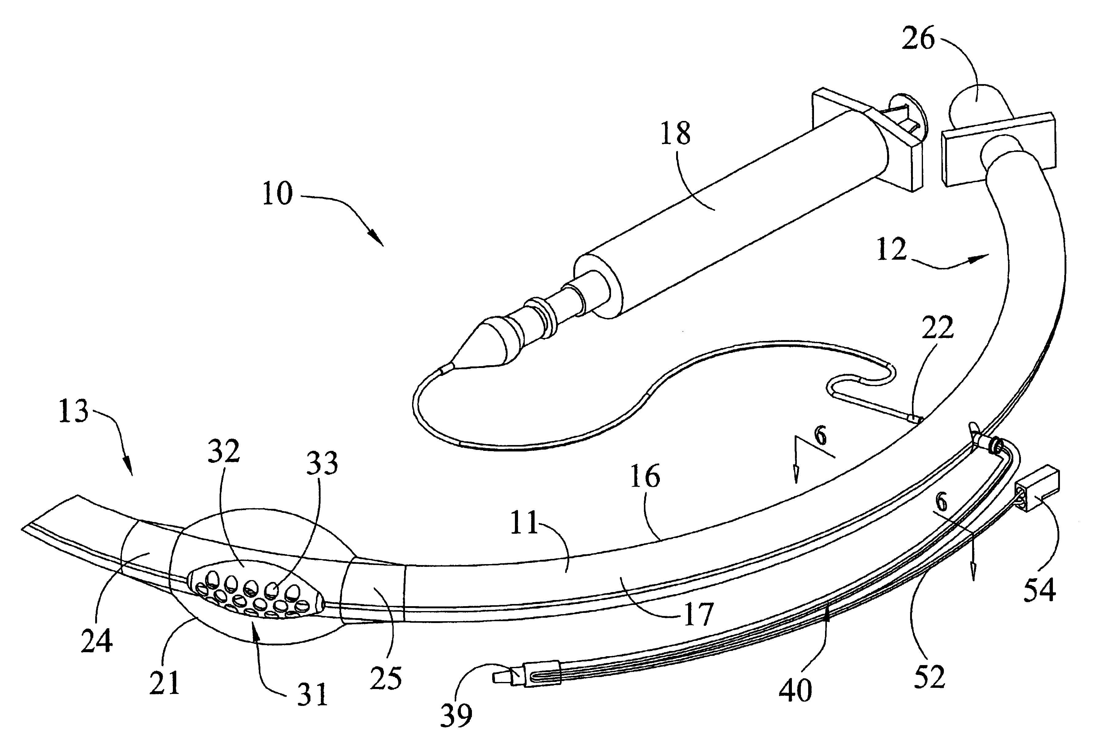
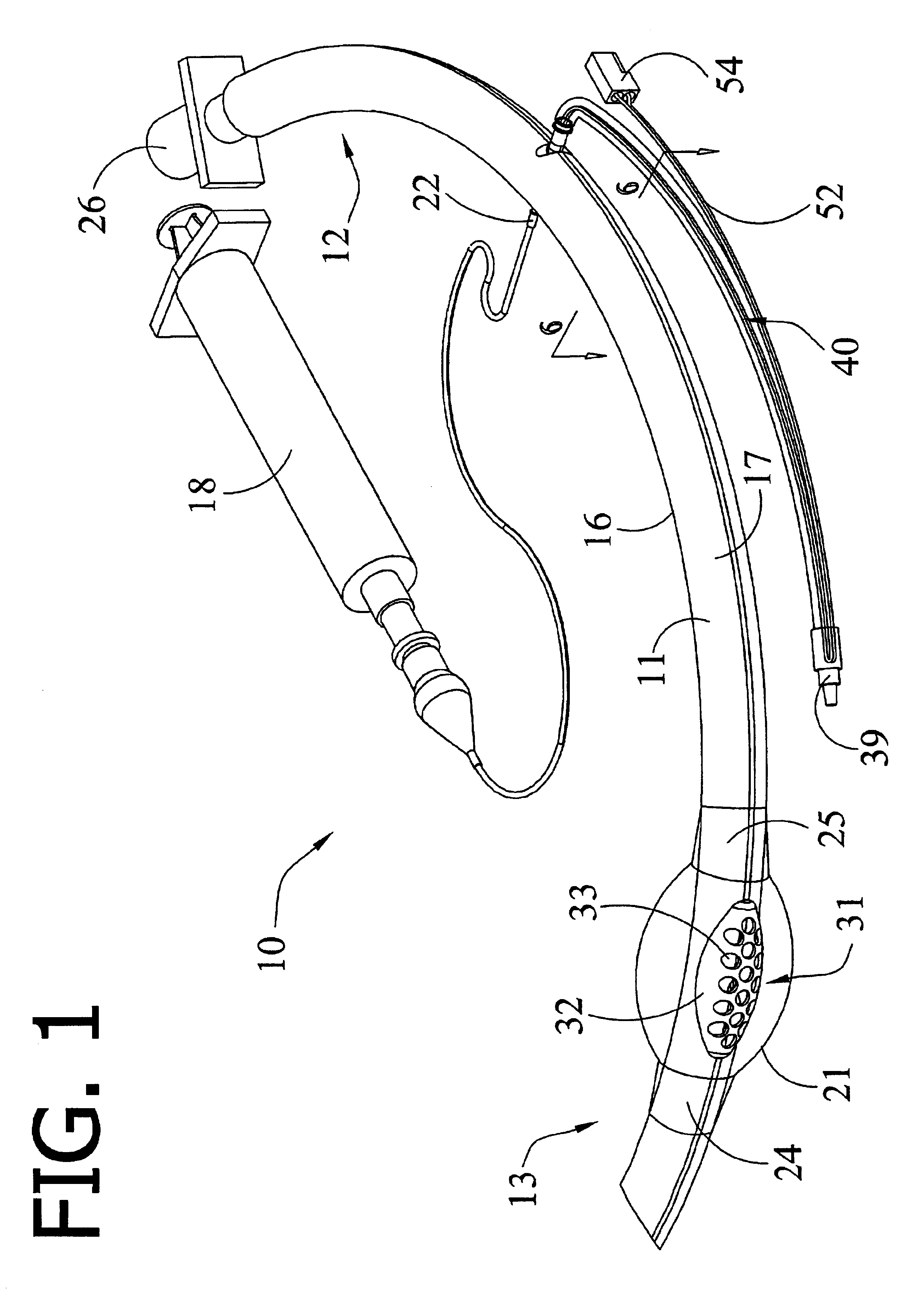
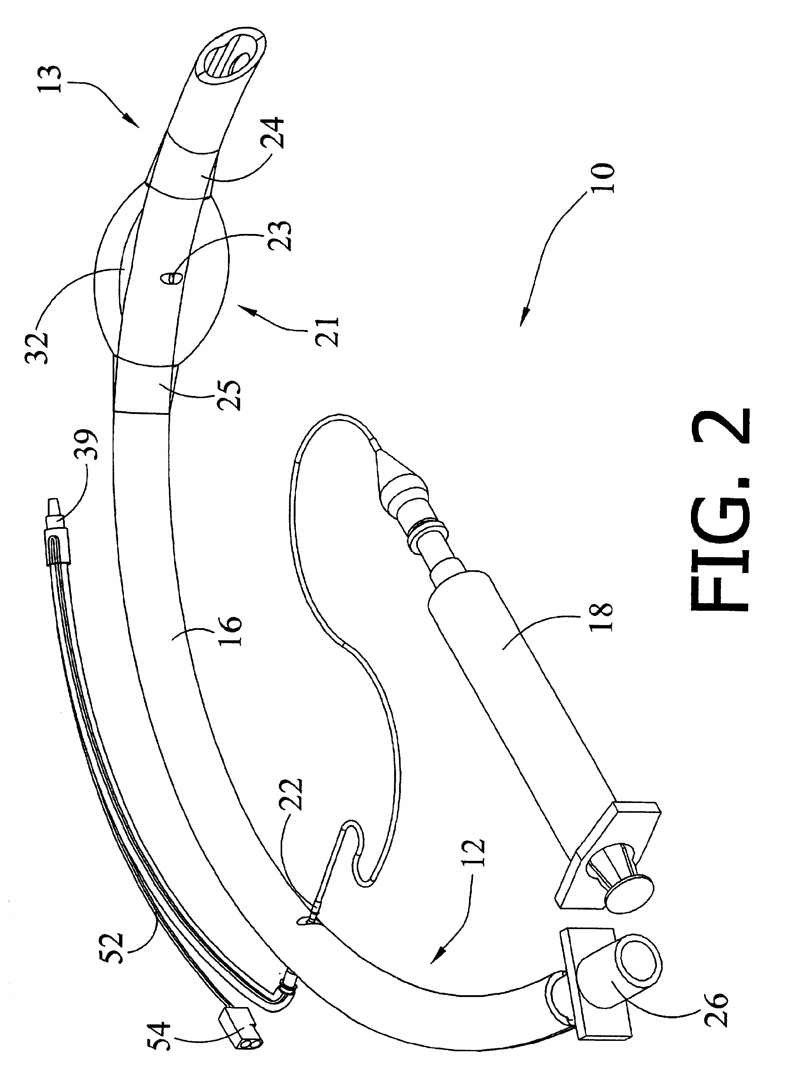
[0023]In reference to the several drawings the multi lumen endotracheal tube 10 of the present invention comprises a longitudinally extending tubular member 11, having a proximal portion 12, and a distal portion 13. Distal portion 13, refers to that portion of ET tube 10 which is inserted ed into a patient's body cavity during intubation, while proximal portion 12 refers to that portion of ET tube 10 which remains external the patient's body. A balloon cuff 21 circumscribing a part of distal portion 13 is selectively inflatable for sealing the trachea. According to the present invention, an ausculatory receiver 25 is provided on distal portion 13 of ET tube 10 to receive a patient's breath sounds. These breath sounds are then transmitted via a fluid media through a tertiary lumen 30 for external monitoring. This arrangement offers health care providers a low cost alternative to electro-optical and electro-audio guided intubation techniques.

[0024]As is known in the art, ET tube 10 is...


PUM


Abstract

A multi lumen endotracheal tube having a balloon cuff to seal a patient's trachea during intubation. The ET tube having a main lumen for the exchange of respiratory and medicinal gases consequent to a medical procedure, a secondary lumen for inflation of the balloon cuff, and a tertiary lumen for transmission of sound waves via air medium contained therein, permitting ausculatory monitoring of a patient's breath sounds during intubation and subsequent monitoring of cardiac and respiratory activity after sealing of the ET tube. The ET tube is particularly a adapted for utilization by an anesthetist by including temperature sensor to permit remote monitoring of body core temperature and body cavity ausculation of cardiac and respiratory activity.

Description

TECHNICAL FIELD[0001]This invention relates to the field of endotracheal tubes having an inflatable cuff to seal a patient's trachea during intubation. More specifically, the device of the present invention is directed to an endotracheal tube adapted for ausculatory monitoring of a patient's breath sounds. With even greater particularity the device may be adapted for utilization by an anesthetist to permit remote monitoring of body core temperature and body cavity ausculation of cardiac and respiratory activity.BACKGROUND OF THE INVENTION[0002]Multi lumen inflatable cuff endotracheal (ET) tubes are well known in the medical arts particularly with respect to anesthesiology. Endotracheal tubes of this type will generally comprise a main lumen to maintain a patient's airway for the passage of respiratory gases to and from the lungs and a secondary lumen for inflation of the balloon cuff after the health care provider has intubated the patient. The inflatable cuff performs various funct...

Claims


Application Information

Patent Timeline
IPC IPC(8): A61F6/02A61F6/00A61M16/04
CPCA61M16/0463A61M16/0434A61M16/044A61M2205/3375A61M2230/04A61M2230/40A61M2230/50Y10S128/911A61M16/0486
Inventor MOORE, JOHNNY V.
Owner MOORE JOHNNY V
Why Eureka
Try Eureka