
Methods and apparatus for accessing and stabilizing an area of the heart

a heart and muscle tissue technology, applied in the field of medical devices and methods for accessing anatomic surfaces, muscle layers, vessels or anatomic space of the body, can solve the problems of unattractive scars, carries additional complications, and thoracic muscle and rib injuries

Inactive Publication Date: 2005-07-19
MEDTRONIC INC
View PDF35 Cites 251 Cited by
  • Summary
  • Abstract
  • Description
  • Claims
  • Application Information

AI Technical Summary

Benefits of technology

[0025]The cutting instrument can be a knife blade, a needle, a stiff guidewire tip, an electrosurgical cutting tool, surgical scissors, or other piercing or cutting tools. Preferably, the cutting instrument comprises a shaped cutting blade having a blade tip and a blade edge that facilitates perforating and cutting a slit through the pericardium to form an elongated pericardial incision.

Problems solved by technology

As a result, these operations typically require large numbers of sutures or staples to close the incision and 5 to 10 wire hooks to keep the severed sternum together.
Such surgery often carries additional complications such as instability of the sternum, post-operative bleeding, and mediastinal infection.
The thoracic muscle and ribs are also severely traumatized, and the healing process results in an unattractive scar.
Therefore, much effort has been expended to develop medical devices and techniques to access the pericardial space employing such minimally invasive percutaneous procedures.
One difficulty has been that normally the pericardial space is so small or thin that it is difficult to penetrate the pericardium using miniaturized instruments capable of being introduced through a port to the site without also puncturing the underling epicardium and thereby, damaging the myocardium or a coronary vessel.
Proliferative adhesions occur between the pericardium and the epicardium in diseased hearts and hamper access to the pericardial space employing such minimally invasive percutaneous procedures.
It is difficult to introduce and use the forceps through the narrow lumen of a port or sleeve, particularly if the pericardial fluid is under pressure that makes the pericardium taut like an inflated balloon.
Although theoretically plausible, the ability to reliably maintain a vacuum seal against the pericardium when such treatment devices are advanced can be problematic.

Method used


Image

  • Methods and apparatus for accessing and stabilizing an area of the heart
  • Methods and apparatus for accessing and stabilizing an area of the heart
  • Methods and apparatus for accessing and stabilizing an area of the heart

Examples


Embodiment Construction

[0074]In the following detailed description, references are made to illustrative embodiments of methods and apparatus for carrying out the invention. It is understood that other embodiments can be utilized without departing from the scope of the invention. Preferred methods and apparatus are described for accessing the pericardial space between the epicardium and the pericardium as an example of accessing an anatomic space between an outer tissue layer and an inner tissue layer.

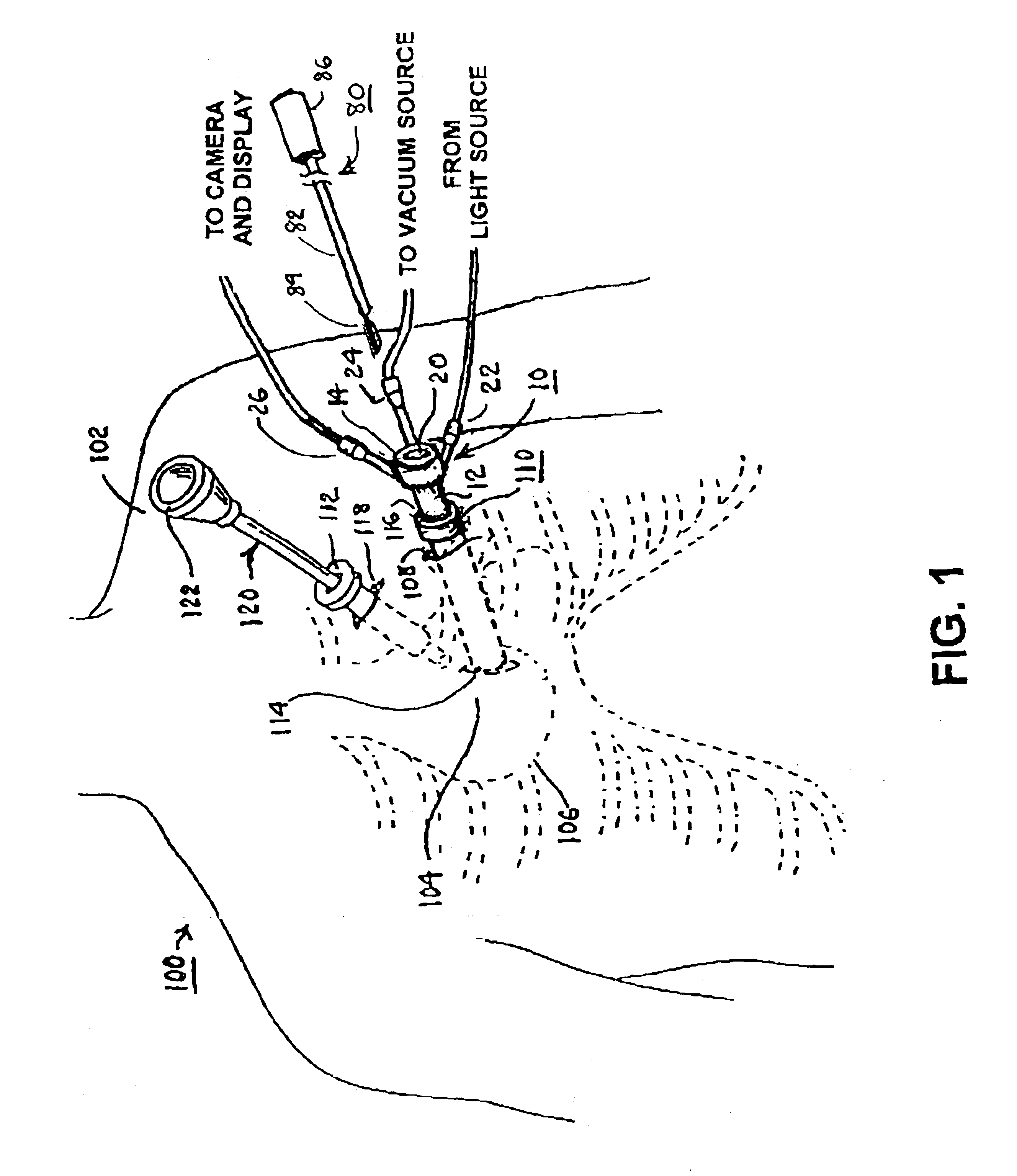
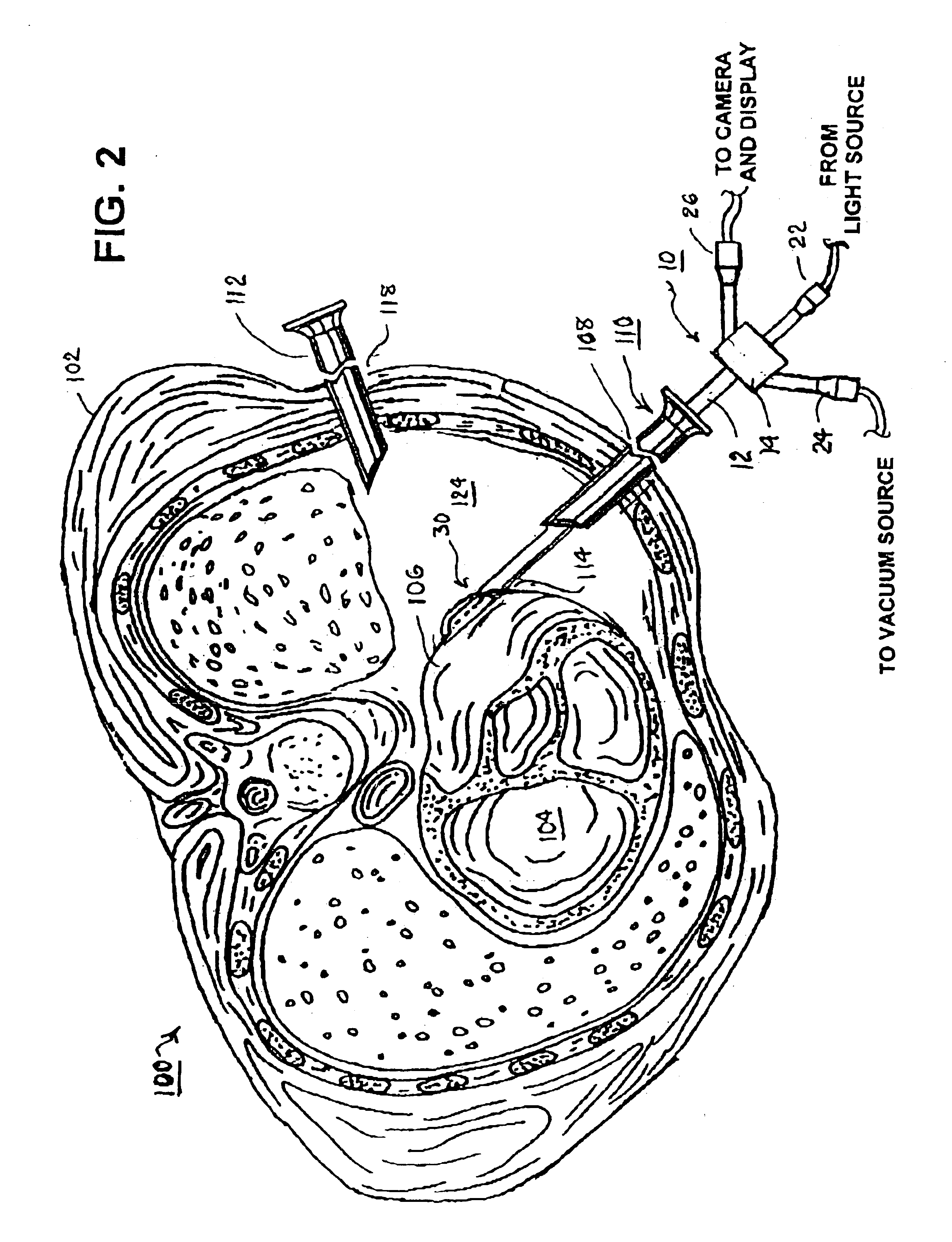
[0075]For example, FIGS. 1-3 illustrate the placement of instruments through the chest wall of a patient 100 for observation and accessing the pericardial space through an incision in the pericardium 106 exposing the pericardium of the heart 104 to perform any of the ancillary procedures listed above. The patient 100 is placed under general anesthesia, and the patient's left lung is deflated if necessary, using conventional techniques. The patient 100 is placed in a lateral decubitus position on his right sid...


PUM


Abstract

A tubular suction tool for accessing an anatomic surface or anatomic space and particularly the pericardium to access pericardial space and the epicardial surface of the heart to implant cardiac leads in a minimally invasive manner are disclosed. The suction tool incorporates a suction pad concave wall defining a suction cavity, a plurality of suction ports arrayed about the concave wall, and a suction lumen, to form a bleb of tissue into the suction cavity when suction is applied. The suction cavity extends along one side of the suction pad, so that the suction pad and suction cavity can be applied tangentially against a tissue site. The suction tool can incorporate light emission and video imaging of tissue adjacent the suction pad. A working lumen terminating in a working lumen port into the suction cavity enables introduction of tools, cardiac leads, and other instruments, cells, drugs or materials into or through the tissue bleb drawn into the suction cavity.

Description

CROSS-REFERENCE TO RELATED APPLICATIONS[0001]This application is a continuation-in-part of U.S. patent application Ser. No. 10 / 342,932 filed Jan. 15, 2003 now U.S. Pat. No. 6,837,848, the disclosure of which is incorporated herein by reference.[0002]This application is related to commonly assigned U.S. patent application Ser. No. 10 / 283,794 filed Oct. 30, 2002, for METHODS AND APPARATUS FOR ACCESSING AND STABILIZING AN AREA OF THE HEART in the names of Gary W. Guenst et al., U.S. patent application Ser. No. 10 / 342,960 filed Jan. 15, 2003, for METHODS AND TOOLS FOR ACCESSING AN ANATOMIC SPACE in the name of Gary W. Guenst, and U.S. patent application Ser. No. 10 / 284,771 filed Oct. 31, 2002, for ANATOMIC SPACE ACCESS SUCTION TOOLS AND METHODS in the names of Koen Michels et al., the disclosures of which are incorporated herein by reference.FIELD OF THE INVENTION[0003]The present invention pertains to medical devices and methods for accessing an anatomic surface, muscle layer, vessel o...

Claims


Application Information

Patent Timeline
Patent Type & Authority Patents(United States)
IPC IPC(8): A61B19/10A61B19/08A61B19/00A61B19/02A61B18/14A61B46/23A61M1/00
CPCA61B18/1485A61B19/08A61B19/10A61M1/008A61B18/1402A61B19/0256A61B2018/00291A61B2019/5206A61B2019/5217A61B2218/007A61B46/23A61B2090/3614A61B50/20A61B46/00A61B2090/306A61M1/84
Inventor BONNER, MATTHEW D.USHER, RAYMOND W.CHEN, VICTOR T.KEOGH, JAMES R.
Owner MEDTRONIC INC
Learn More