

Analysis of fundus images
a fundus image and image technology, applied in image data processing, instruments, othalmoscopes, etc., can solve the problems of higher structure complexity, achieve good estimation of the position of the papilla, and improve the accuracy of detection results
- Summary
- Abstract
- Description
- Claims
- Application Information
AI Technical Summary
Benefits of technology
Problems solved by technology
Method used
Image

Examples
Embodiment Construction
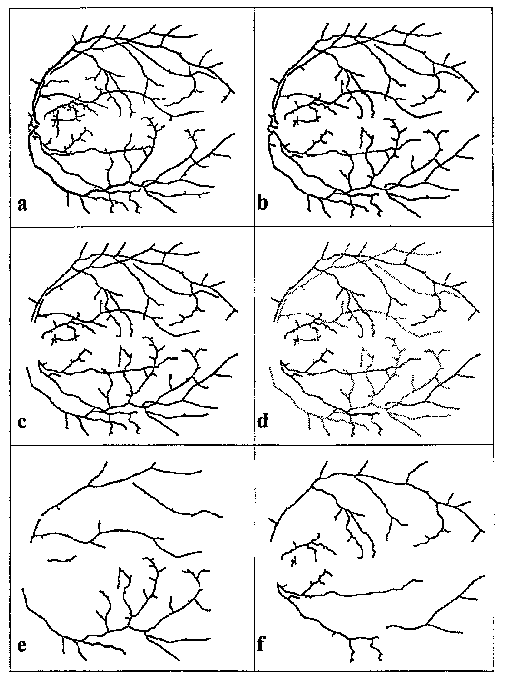
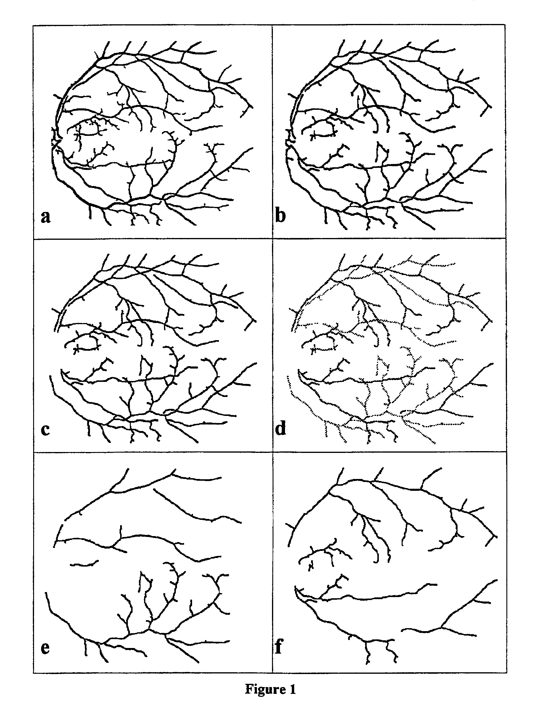
[0050]A number of preferred embodiments of the invention will now be described with reference to the accompanying FIG. 1, which is a graphical representation of vascular structure in a digitized ocular fundus image, and illustrates how the arterial and venous systems may be derived from the image.
[0051]Finding the Arteriolar and Venous Systems
[0052]Various methods are known by which the vascular system may be isolated from the rest of the image content and skeletonized. For example a method may be employed based on the one described in the article by Subhasis Chaudhuri et al, “Detection of Blood Vessels in Retinal Images Using Two-Dimensional Matched Filters”, IEEE Transactions on Medical Imaging, Vol. 8, No. 3, September 1989. In this method, use is made of the fact that the vessels are linear in a local neighborhood, where different filter matrices have different orientations. The localization and orientation of such line elements may be determined using a template matching approa...
PUM
 Abstract
Description
Claims
Application Information