Suturing device and method for sealing an opening in a blood vessel for other biological structure

a suturing device and blood vessel technology, applied in the field of medical suturing devices, can solve the problems of thrombosis, thrombosis bursting, patient bleeding to death, etc., and achieve the effect of reducing complications and costs

Inactive Publication Date: 2006-02-28
WHITEBOX CAPITAL ARBITRAGE PARTNERS +1
View PDF44 Cites 214 Cited by
  • Summary
  • Abstract
  • Description
  • Claims
  • Application Information

AI Technical Summary

Benefits of technology

[0011]The present invention addresses the above problems by providing a suturing device and method for suturing an opening in a biological tissue wall, such as an organ or blood vessel. The device is particularly well suited to suture an opening made in an artery, such as the femoral artery, following a catheterization procedure. The device eliminates the need to apply pressure to a patient's thigh for an extended period of time, and eliminates many of the complications and costs associated with the creation of a thrombus patch.

Problems solved by technology

A significant problem with this approach is that it is frequently necessary to apply the pressure for an extended period of time, such as twenty-four hours or longer.
Another problem with the thrombus patch method is that the high blood pressure in the artery can cause the thrombus patch to rupture or burst while direct pressure is being applied to the thigh or after direct pressure is removed.
If the patch ruptures and is not restored, the patient may bleed to death.
Because thrombus patches frequently burst, the patient frequently must remain in the hospital or catheterization lab overnight for observation.
Thus, these ‘out-patient’ procedures become ‘in-patient’ procedures, simply because a thrombus patch it is difficult to create.
Staying in the hospital increases patient discomfort and hospital expenses, which are often disproportionate to the actual medical procedure performed.
Furthermore, if a thrombus patch cannot be formed, the physician may need to anesthetize the patient, occlude blood flow to the artery, make a large incision in the thigh to allow conventional suturing with a needle, suture the artery with conventional means, restore blood flow to the artery, and suture the incision in the thigh.
This results in additional discomfort and expenses for the patient.
While the above problems could potentially be avoided by suturing the blood vessel immediately following the catherization procedure, the size and location of the artery make suturing difficult.
Specifically, the opening in the thigh is typically too small and too deep to provide enough working space for suturing the artery using conventional methods.
Thus, in order to suture the vessel according to conventional methods, the opening in the thigh would have to be significantly enlarged, potentially exposing the patient to additional pain, scarring, and health risks.

Method used


Image

  • Suturing device and method for sealing an opening in a blood vessel for other biological structure
  • Suturing device and method for sealing an opening in a blood vessel for other biological structure
  • Suturing device and method for sealing an opening in a blood vessel for other biological structure

Examples


Embodiment Construction

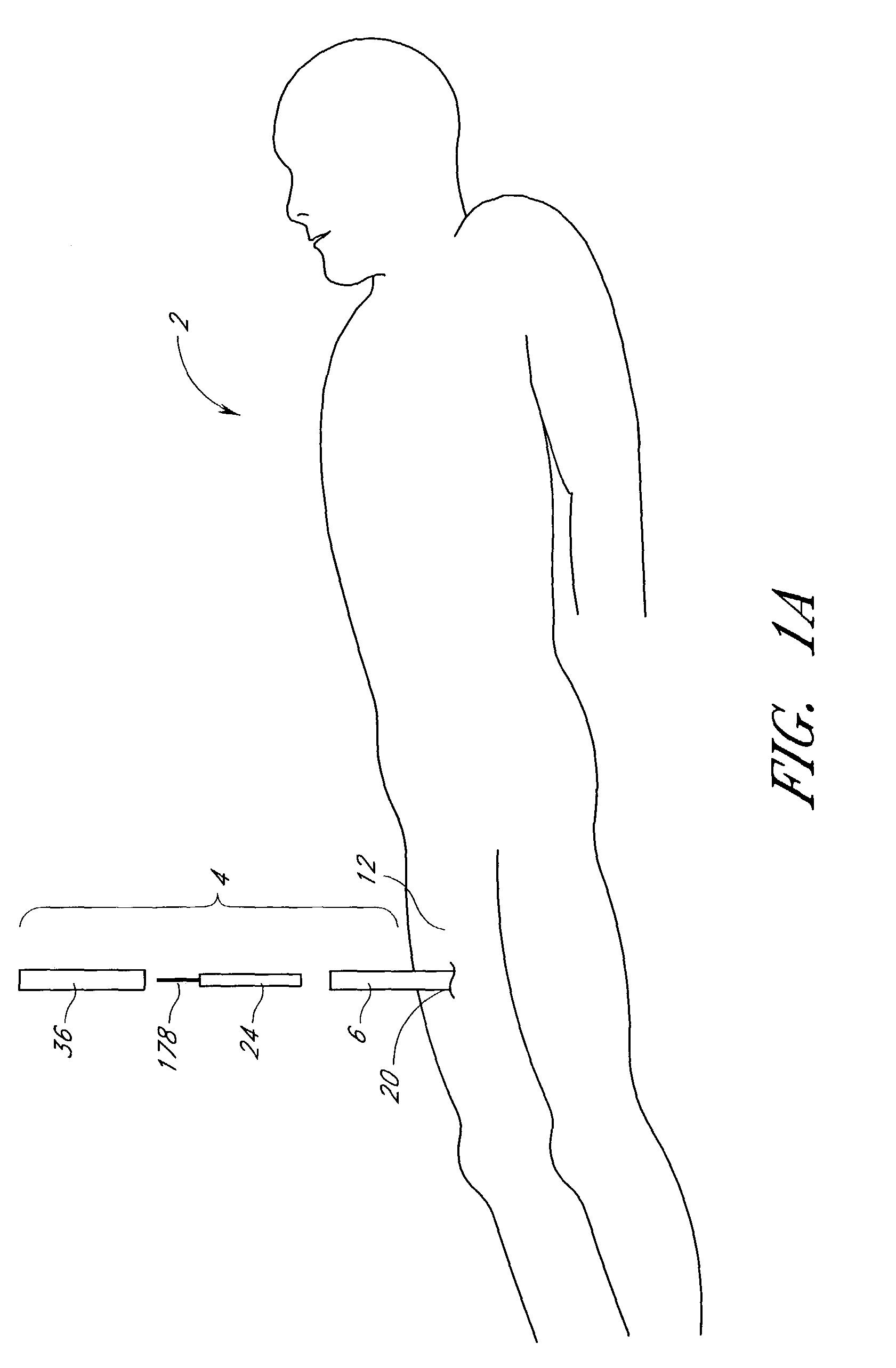
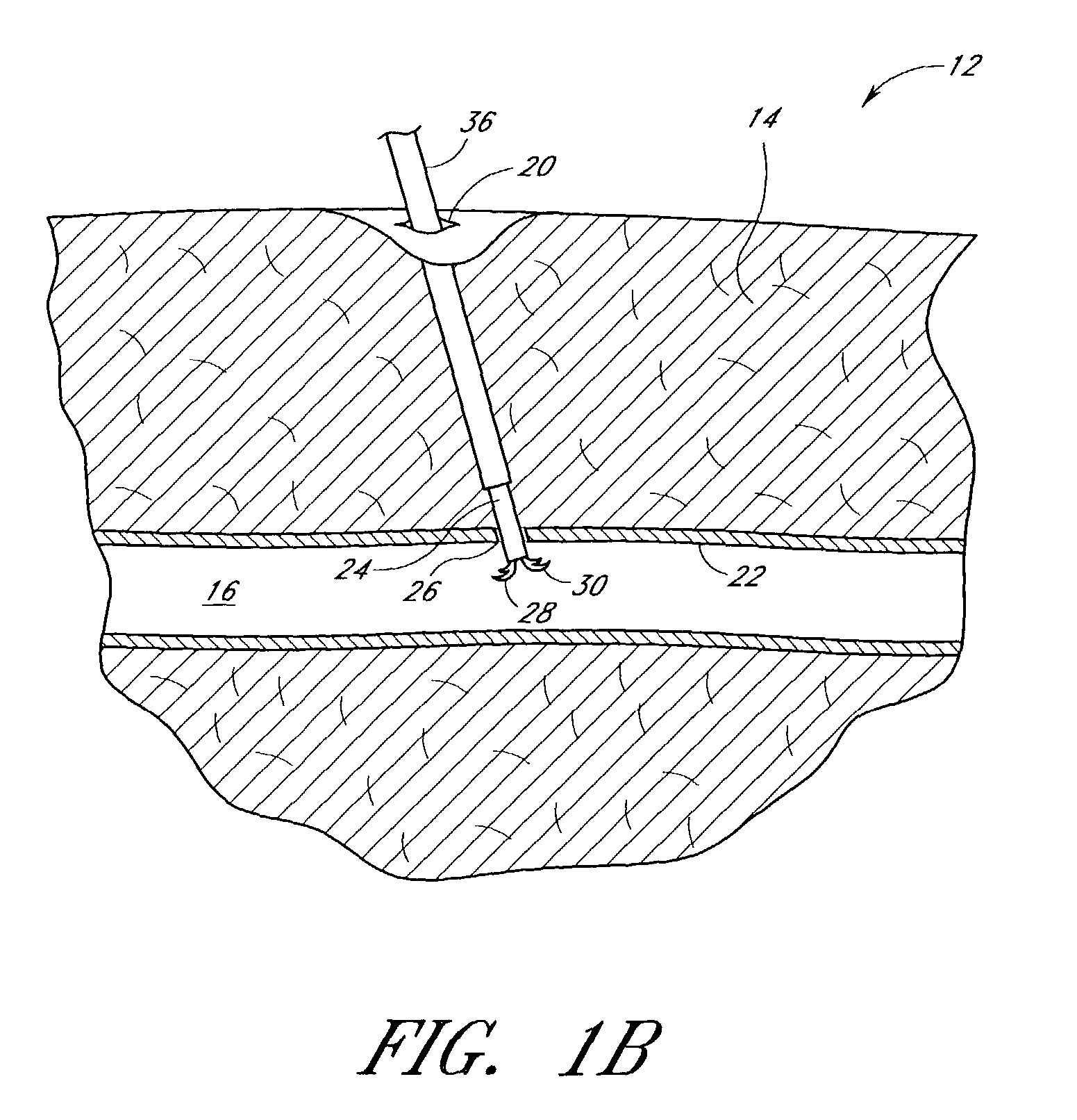
[0083]The present invention provides a suturing device for suturing biological tissue. The suturing device may be used to seal a blood vessel following an interventional catheterization procedure, such as an angiogram. FIGS. 1A–1B illustrate one embodiment of the present invention in an exemplary use environment. As depicted by FIGS. 1A–1B, the physician makes an initial incision 20 in the upper thigh 12 of a patient 2. The physician then inserts a needle (not shown) into the incision 20. When blood bleeds back from the insertion, the physician knows the needle has pierced the femoral artery 16. The physician then inserts a guidewire (not shown) through the needle and into the artery. The physician may take the needle out and insert a plastic needle (not shown) over the guidewire once the guidewire is in place. The guidewire may then be taken out.

[0084]With this needle in place, the physician can insert a catheter sheath introducer (CSI) 6, also called an introducer sheath. This int...


PUM


Abstract

A suturing device allows a physician to remotely seal an incision in a blood vessel or other biological tissue. The device comprises an elongated tubular body having a distal portion which is adapted to be inserted percutaneously through the incision and into the blood vessel. The distal portion has first and second retractable arms which extend from the distal portion of the body and releasably hold a suture within the blood vessel. First and second retractable needles, each of which is configured to catch the suture from a respective arm, are provided along the body proximal to the retractable arms. The arms and the needles are remotely movable by the physician using a handle or other control mechanism provided at a distal portion of the device. In operation, the arms are initially deployed within the blood vessel to hold the ends of the suture beyond the circumference of the tubular body. The needles are then deployed from and then retracted into the body, during which time the needles pierce the vessel wall on opposite sides of the incision, release the suture ends from the retractable arms, and pull the suture through the vessel wall. The device is particularly useful for closing an incision in an artery following a catheterization procedure. In one embodiment, the catheter sheath introducer (CSI) used to perform the catheterization procedure is left in place during the suturing procedure.

Description

RELATED APPLICATIONS[0001]This application is a continuation of U.S. application Ser. No. 09 / 773,046 filed Jan. 30, 2001 now U.S. Pat. No. 6,551,331, which is a continuation of U.S. application Ser. No. 09 / 471,866, filed Dec. 23, 1999 now U.S. Pat. No. 6,245,079, which is a continuation of U.S. application Ser. No. 09 / 231,177, filed Jan. 14, 1999, now U.S. Pat. No. 6,117,144, which is a continuation-in-part of U.S. application Ser. No. 09 / 036,437, filed Mar. 9, 1998, now abandoned, which is a continuation-in-part of application Ser. No. 08 / 702,315 filed Aug. 23, 1996, now U.S. Pat. No. 5,860,990, which claims the benefit of U.S. Provisional Application No. 60 / 002,769 filed Aug. 24, 1995.BACKGROUND OF THE INVENTION[0002]1. Field of the Invention[0003]The present invention relates to medical suturing devices. More particularly, the present invention relates to suturing devices for closing an opening in an arterial or other biological tissue wall that is not directly accessible to the ...

Claims


Application Information

Patent Timeline
Patent Type & Authority Patents(United States)
IPC IPC(8): A61B17/04A61B17/00A61B17/06A61B17/28
CPCA61B17/0057A61B17/0469A61B17/0482A61B17/0483A61B17/0491A61B2017/2911A61B2017/00663A61B2017/00672A61B2017/00867A61B2017/0472A61B2017/06042A61B2017/00637
Inventor NOBLES, ANTHONY A.DECKER, STEVEN E.BROSCH, BENJAMIN G.
Owner WHITEBOX CAPITAL ARBITRAGE PARTNERS
Why Eureka
Try Eureka