Spatial-temporal lesion detection, segmentation, and diagnostic information extraction system and method

a technology of diagnostic information and detection system, applied in the field of spatial-temporal lesion detection, segmentation, and diagnostic information extraction system and method, can solve the problems of complex process of diagnosis of lesion/cancer, unable to achieve clinically significant results, and unable to achieve medical diagnosis based on such data,

Active Publication Date: 2006-08-08
EDDA TECH MEDICAL SOLUTIONS SUZHOU LTD +1
View PDF5 Cites 54 Cited by
  • Summary
  • Abstract
  • Description
  • Claims
  • Application Information

AI Technical Summary

Problems solved by technology

The process of diagnosing a lesion / cancer is complex and often requires consideration of different types of diagnostic information extracted from different sources such as diagnostic images.
Even with such information afforded by modern technologies, diagnosis of lesion / cancer in some organs such as liver lesion / cancer has still proven to be a significant challenge.
Given those difficulties in processing data from various modalities, reaching a medical diagnosis based on such data often requires substantial clinical experience.

Method used


Image

  • Spatial-temporal lesion detection, segmentation, and diagnostic information extraction system and method
  • Spatial-temporal lesion detection, segmentation, and diagnostic information extraction system and method
  • Spatial-temporal lesion detection, segmentation, and diagnostic information extraction system and method

Examples


Embodiment Construction

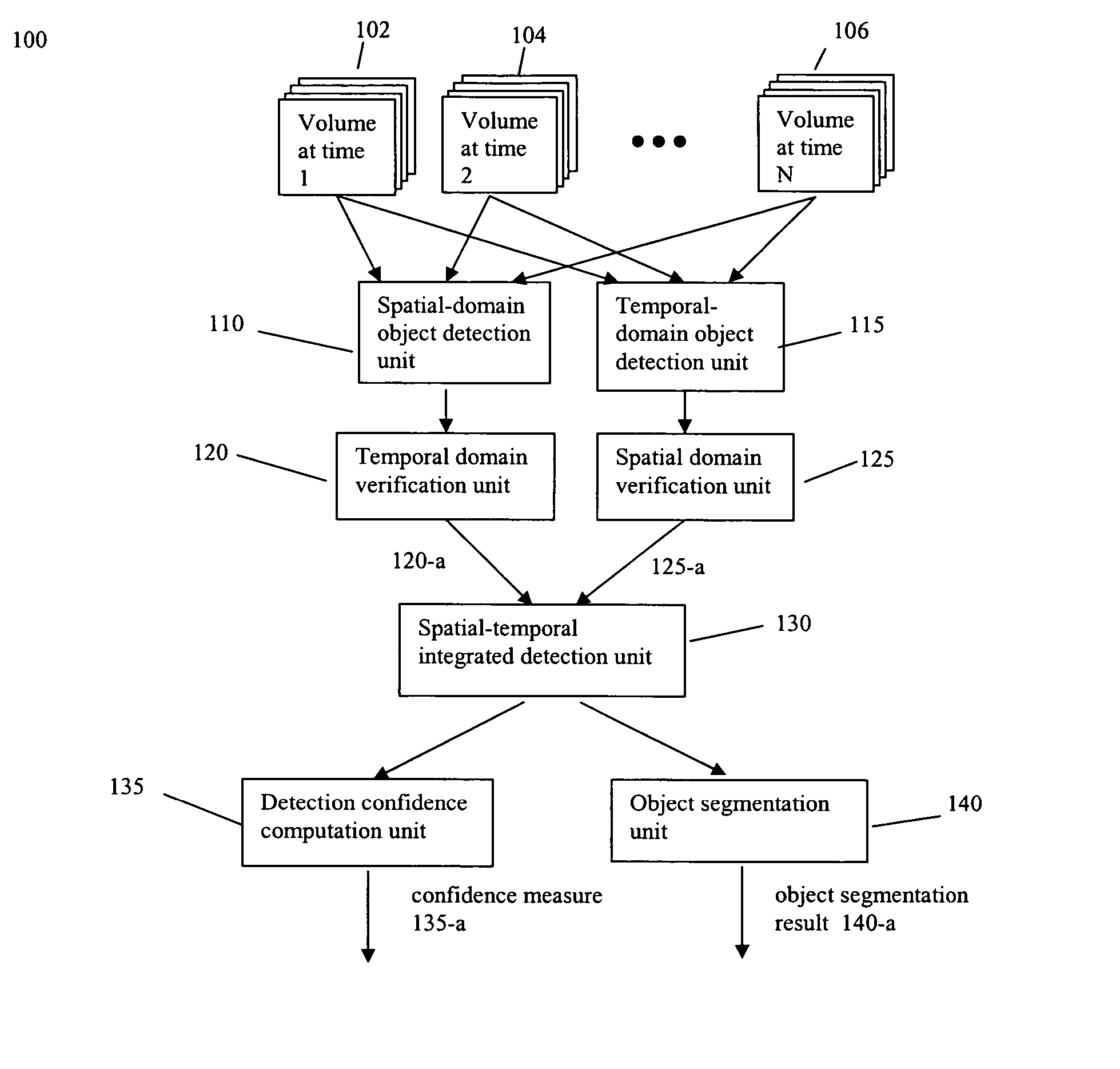
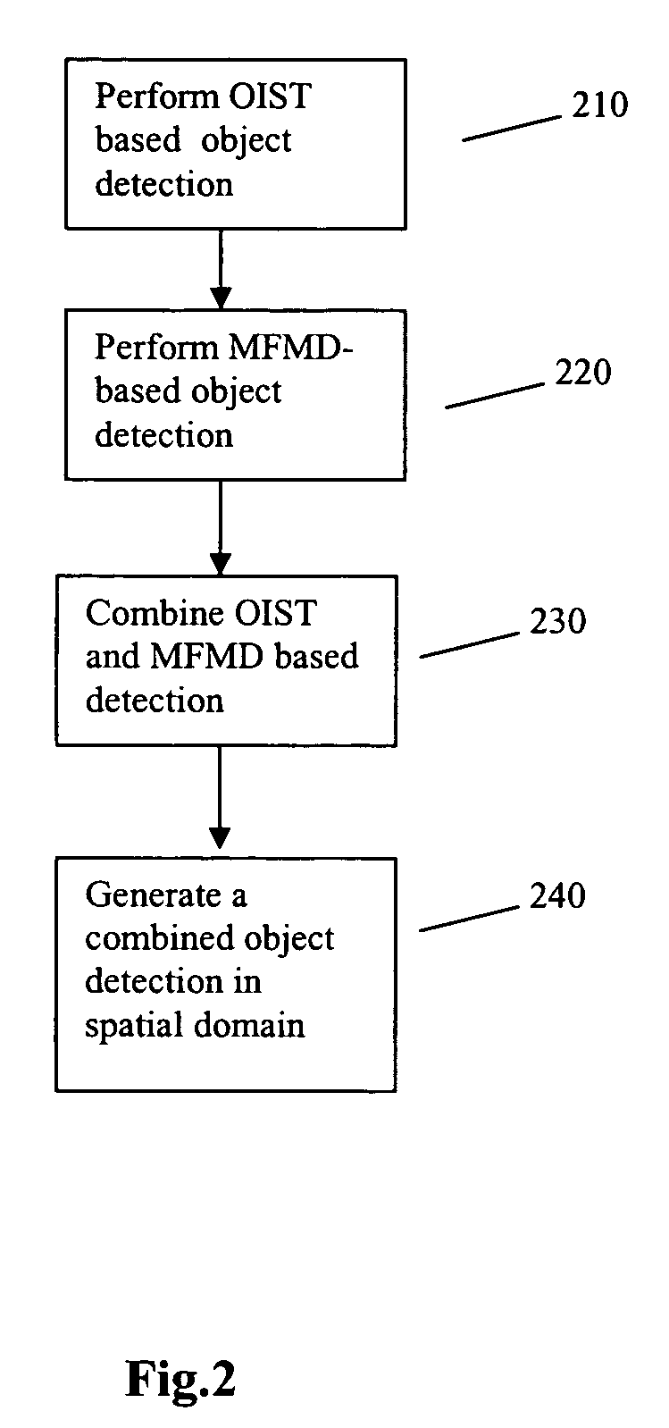
[0018]The present invention relates to methods and systems for lesion detection, segmentation, and feature extraction. Methods and systems are disclosed herein that enable integrated spatial temporal processing of imagery information facilitating identification of a lesion contained therein and quantification thereof. In some embodiments, an OIST (organ intensity subtraction) image derived from an image may be used to detect a lesion with a homogeneous intensity distribution. For example, when an organ under a patient study is a liver, then a liver intensity subtraction (LIST) image may be obtained from an image containing a liver. An MFMD map derived from the same image may be used to detect a lesion with an inhomogeneous intensity distribution. An OIST based detection result may be combined with an MFMD based detection result to generate an integrated spatial domain lesion detection result. In some embodiments, a lesion may also be detected in the temporal domain based on an organ...


PUM


Abstract

Method and system for lesion detection is disclosed. A lesion is detected from each data set in the spatial domain based on an organ intensity subtracted (OIST) image. In the temporal domain, the lesion is detected based on a maximum frequency-magnitude difference (MFMD) map across a plurality of data sets. The detection result from the spatial domain and the detection result from the temporal domain are integrated to produce an integrated lesion detection result.

Description

[0001]The present invention claims priority of provisional patent application No. 60 / 562,259 filed Apr. 15, 2004, the contents of which are incorporated herein in their entirety.BACKGROUND OF THE INVENTION[0002]1. Field of the Invention[0003]The present invention relates generally to a system and method for medical image processing. Specifically, the present invention relates to a method and system for computer assisted medical diagnosis and systems incorporating the present invention.[0004]2. Description of Related Art[0005]The process of diagnosing a lesion / cancer is complex and often requires consideration of different types of diagnostic information extracted from different sources such as diagnostic images. Such diagnostic images may be acquired in different modalities such as Ultrasound (US), Computerized Tomography (CT), or Magnetic Resonance Imaging (MRI). Each image modality may offer different insight regarding a disease and provide distinct medical evidence that can be us...

Claims


Application Information

Patent Timeline
Patent Type & Authority Patents(United States)
IPC IPC(8): G06K9/00G06K9/32G06T5/00G06T7/00
CPCG06T7/0012G06T7/0081G06T7/0095G06T7/0097G06T2207/30056G06T2207/10132G06T2207/20056G06T2207/20156G06T2207/30004G06T2207/10072G06T7/11G06T7/168G06T7/174
Inventor WEI, GUO-QINGQIAN, JIAN-ZHONGFAN, LILIANG, CHENG-CHUNG
Owner EDDA TECH MEDICAL SOLUTIONS SUZHOU LTD
Why Eureka
Try Eureka