Automatic analysis in virtual endoscopy

Inactive Publication Date: 2006-12-12
WAKE FOREST UNIVERSITY
View PDF75 Cites 237 Cited by
  • Summary
  • Abstract
  • Description
  • Claims
  • Application Information

AI Technical Summary

Benefits of technology

[0009]In accordance with the present invention, a method and system are provided for interactively displaying a three-dimensional rendering of a structure having a lumen wherein the rendering is effected by a an adaptive thresholding procedure. A three-dimensional volume of data is formed in a computer from a series of two-dimensional images acquired, for example, by a scanner such as CT-scanner. The acquired images represent at least one physical property associated with the three-dimensional object. A selected region of interest is segmented by the computer from the vo

Problems solved by technology

However, only about 1.5 million fiberoptic colonoscopies are performed each year.
Another problem is that conventional colonoscopies fail to provide access to the entire colon in approximately 15% of

Method used


Image

  • Automatic analysis in virtual endoscopy
  • Automatic analysis in virtual endoscopy
  • Automatic analysis in virtual endoscopy

Examples


Embodiment Construction

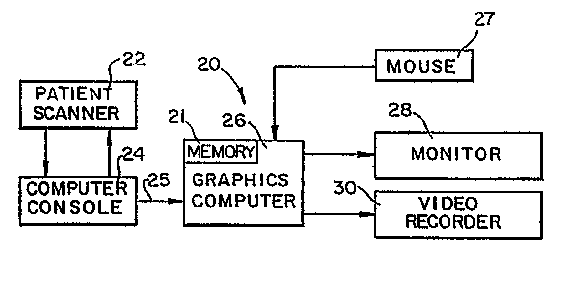
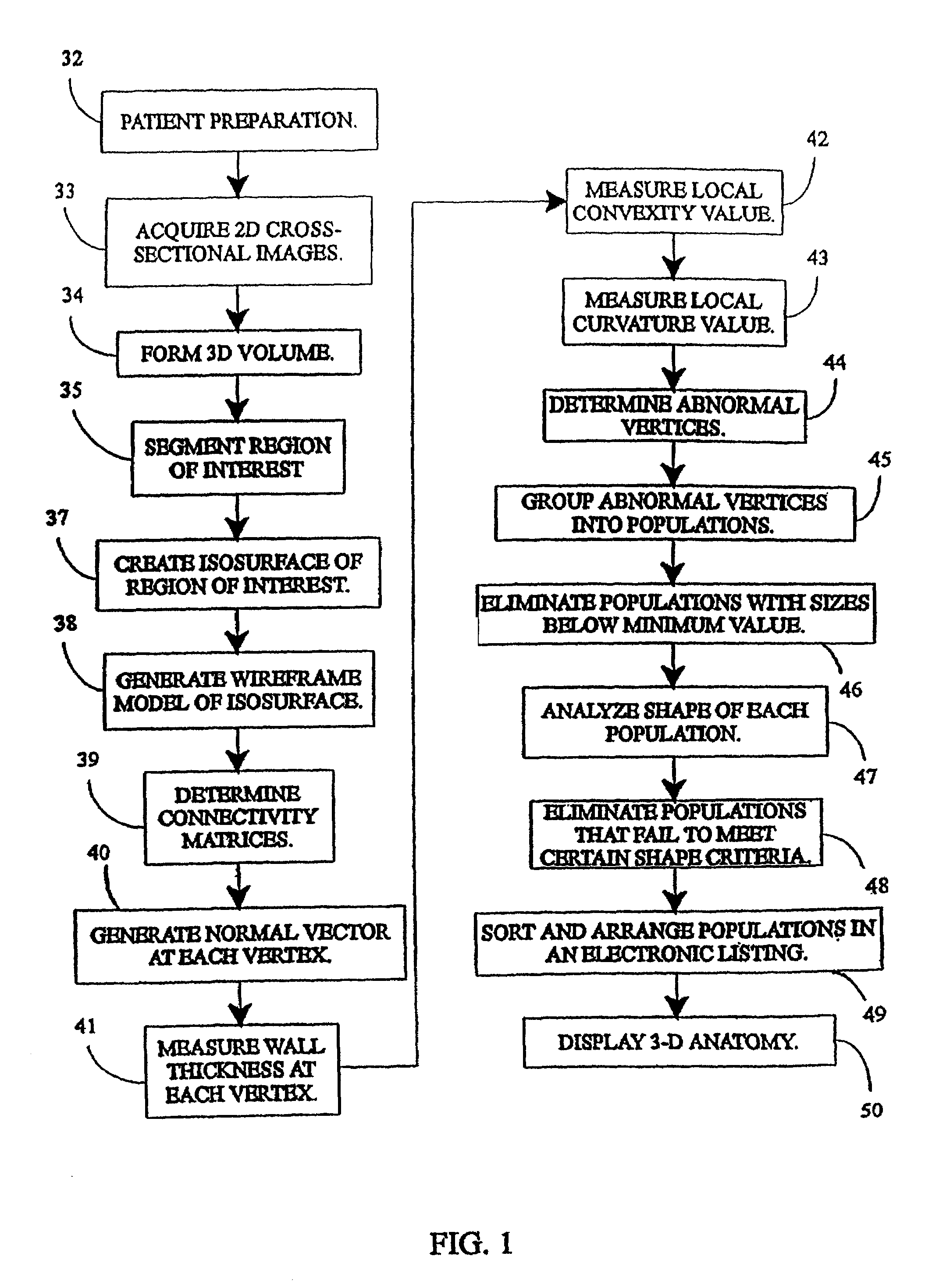
[0021]The present invention generally relates to a method and system, as schematically represented in FIGS. 1–4, for generating interactive, three-dimensional renderings of three-dimensional structures generally having a lumen. The structures are usually in the general form of selected regions of a body and in particular, human or animal body organs which have hollow lumens such as colons, blood vessels, and airways. In accordance with the present invention, the interactive three-dimensional renderings are generated in a computer-controlled process from a series of two-dimensional, cross-sectional images of the selected body organ acquired, for example, from a helical (computed tomography (CT) Scan. The three-dimensional renderings are interactive in that such renderings can be manipulated by a user on a visual display of a computer system, such as a computer monitor, to enable movement in, around and through the three-dimensional structure while simultaneously displaying multiplana...


PUM


Abstract

A computer system and a computer-implemented method are provided for interactively displaying a three-dimensional rendering of a structure having a lumen and for indicating regions of abnormal wall structure. A three-dimensional volume of data is formed from a series of two-dimensional images representing at least one physical property associated with the three-dimensional structure. An isosurface of a selected region of interest is created by a computer from the volume of data based on a selected value or values of a physical property representing the selected region of interest. A wireframe model of the isosurface is generated by the computer wherein the wireframe model includes a plurality of vertices. The vertices are then grouped into populations of contiguous vertices having a characteristic indicating abnormal wall structure by the computer. The wireframe model is then rendered by the computer in an interactive three-dimensional display to indicate the populations of abnormal wall structure.

Description

RELATED APPLICATION[0001]This application is a continuation of application Ser. No. 09 / 299,061, entitled “Automatic Analysis in Virtual Endoscopy”, filed on Apr. 23, 1999, now issued as U.S. Pat. No. 6,366,800, which in turn is a continuation of application Ser. No. 08 / 805,584, entitled “Automatic Analysis in Virtual Endoscopy”, filed on Feb. 25, 1997, now issued as U.S. Pat. No. 5,920,319, which in turn is a continuation-in-part of application Ser. No. 08 / 331,352, filed on Oct. 27, 1994, now issued as U.S. Pat. No. 5,782,762, which are each incorporated herein by reference.FIELD OF THE INVENTION[0002]The present invention relates to a system and method for interactively displaying a three-dimensional rendering of a structure having a lumen and, more particularly, to a system and method for automatically analyzing such structures for potential abnormalities.BACKGROUND OF THE INVENTION[0003]For many forms of cancer, early detection is essential for a favorable prognosis. A cancerous ...

Claims


Application Information

Patent Timeline
IPC IPC(8): A61B5/00A61B1/04A61B5/107A61B6/03G06T1/00G06T5/00G06T15/08G06T17/00G06T17/20G09B23/28
CPCA61B5/1076G06T7/0012G06T7/0081G06T7/0091G06T15/08G06T17/00G06T17/20G09B23/285G06T2207/10081G06T2207/20012G06T2207/30032G06T2207/30064Y10S128/92G06T7/11G06T7/155Y02A90/10
Inventor VINING, DAVID J.HUNT, GORDON W.AHN, DAVID K.STELTS, DAVID R.GE, YAORONGHEMLER, PAUL F.SALIDO, TIFFANY W.
Owner WAKE FOREST UNIVERSITY
Why Eureka
Try Eureka